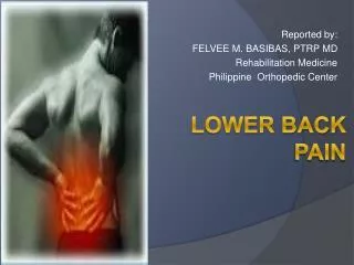
LOWER BACK PAIN
Reported by: FELVEE M. BASIBAS, PTRP MD Rehabilitation Medicine Philippine Orthopedic Center . LOWER BACK PAIN. LOW BACK PAIN. A symptom, not a disease Generally described as pain between the costal margin and the gluteal folds. Extremely common

LOWER BACK PAIN
E N D
Presentation Transcript
Reported by: FELVEE M. BASIBAS, PTRP MD Rehabilitation Medicine Philippine Orthopedic Center LOWER BACK PAIN
LOW BACK PAIN • A symptom, not a disease • Generally described as pain between the costal margin and the gluteal folds. • Extremely common • The leading cause of disability and loss of productivity.
LUMBAR SPINE • Has a dichotomous role in terms of function; • Strength • Flexibility • Performs a major role in support and protection of the spinal contents • Gives us inherent flexibility.
Results from the following: • Size and arrangement of the bones • Arrangement of the ligaments and muscles. • Results from the large number of joints placed so closely together in series (typical lordotic framework). • Also increases ability to absorb shock. STRENGTH FLEXIBILITY
The LUMBAR SPINE • Five lumbar vertebrae • Small percentage has four (sacralization of L5) or six (lumbarization of S1).
LUMBAR VERTEBRA • Components: • Vertebral body • Neural arch • Posterior elements • The vertebral bodies increase in size caudally. • Lower 3 are more wedge shaped (taller anteriorly): creates normal lumbar lordosis. • Serves as weight-bearing function.
Sides of the bony neural arch • Thick pillars that connect the posterior elements to the vertebral body • Resist bending • Transmit forces back and forth from the vertebral bodies to the posterior elements. PEDICLES
Components: • Laminae • Articular processes • Spinous processes • Zygapophyseal joints: created by the superior and inferior articular processes of adjacent vertebrae • Pars interarticularis: • a part of the lamina between the superior and inferior articular processes. • The site of stress fractures (spondylosis), because it is subjected to large bending forces. POSTERIOR ELEMENTS
IV Disk and its attachment to the vertebral end plate are considered secondary cartilaginous joint, or symphysis. • Main function: shock absorption • Annulus fibrosus: acts as the primary shock absorber. INTERVERTEBRAL DISK
NUCLEUS PULPOSUS: • Geletinous inner section of the disk • Consists of water, proteoglycans and collagen. • At birth- 90% water • Desiccate and degenerate as we age. • ANULUS FIBROSUS: • Consists of concentric layers of fibers at oblique angles to each other • Withstand strains in any direction • Outer fibers: more collagen and less proteoglycans and water • Acts more as a ligament to resist flexion, extension, rotation and distraction forces.
BIOMECHANICS Flexion of the lumbar spine Nucleus pulposus is displaced posteriorly Herniation thru the posterior annular fibers (posterolateral disk herniations)
The posterolateral portion of the disk is most at risk, with forward flexion accompanied by lateral bending (i.e. bending and twisting). • Increase in torsional shear forces once the zygapophyseal joints can no longer resist rotation of the lumbar spine; most risky for lumbar disks.
2 sets: • Longitudinal ligaments • Anterior longitudinal ligament (2x stronger) • Posterior longitudinal ligament • Segmental ligaments • Ligamentum flavum • Supraspinous – resist flexion • Interspinous • Intertransverse • Muscles with origin on the lumbar spine • Abdominal musculature • Thoracolumbar fasciae • Pelvic stabilizers THE LIGAMENTS THE MUSCLES
Biomechanical Lifting in Relation to Muscular Activity and Disk Loads • When the muscles contract, there’s associated rise in disk pressure. • These change in pressures depend on the spine posture and the activity undertaken. • There is no significant difference in disk pressure when lifting with the legs (i.e. with the back straight and knees bent) versus lifting with the back (i.e. with a forward-flexed back and straight legs.)
What decreases the forces on the lumbar spine is lifting the load close to your body, as the farther the load is from the chest, the greater the stress on the lumbar spine.
HISTORY • 85% of patients- no specific cause for LBP. • 85% of a diagnosis is made using history alone • Know the following: • Features (location, character, severity, timing) • Alleviating and aggravating factors • Associated signs and symptoms.
RED FLAGS of LBP • Back pain in children <18 years old with considerable pain, or onset in those >55 years old. • History of violent trauma • Constant progressive pain at night • History of CA • Systemic steroids • Drug abuse, HIV infection • Weight loss • Systemic illness
RED FLAGS of LBP • Persisting severe restriction of motion • Intense pain with minimal motion • Structural deformity • Difficulty with micturation • Loss of anal sphincter tone or fecal incontinence, saddle anesthesia • Widespread progressive motor weakness or gait disturbance • Inflammatory disorders (ankylosing spondylitis) suspected
RED FLAGS of LBP • Gradual onset, <40 years • Marked morning stiffness • Persisting limitation of motion • Peripheral joint involvement • Iritis, skin rashes, colitis, urethral discharge • Family history
YELLOW FLAGS of LBP • Signs that the patient who is experiencing low back pain needs further psychologic evaluation. • That the clinician should proceed with caution. • Psychosocial factors
YELLOW FLAGS of LBP • The presence of catastrophic thinking • Expectations that the pain will only worsen with work or activity • Behaviors such as avoidance of normal activity, and extended rest. • Poor sleep • Compensation issues • Emotions such as stress and anxiety • Work issues such as poor job satisfaction • Extended time of work
PHYSICAL EXAMINATION • A. OBSERVATION • Skin, muscle mass, and bony structures. • Posture • Position of lumbar spine • Gait
B. PALPATION • Should begin superficially and progress to deeper tissues • Prone stability testing: • Pressure over isolated vertebrae is applied to look for painful level.
C. RANGE OF MOTION • Quantity of ROM • Single or double inclinometer. • The distance of fingertips to floor • Schober’s test • DOUBLE INCLINOMETER • Correlate the closest to measurements on radiographs • Quality of ROM
D. NEUROLOGIC EXAMINATION • Look for the following: • Weakness • Sensory loss • Diminished/absent reflexes • Special tests – SLR • E. ORTHOPEDIC SPECIAL TESTS TO ASSESS FOR RELATIVE STRENGTH • Curl Trunk Sit Up • Holding the low back flat during lowering
F. ORTHOPEDIC SPECIAL TESTS FOR LUMBAR SEGMENTAL INSTABILITY • Segmental instability – responds to specific stabilization treatment. • SPECIAL TESTS: • PASSIVE INTERVERTEBRAL MOTION TESTING • Prone position • Pressure over spinous process • Assess: amount of vertebral motion and if pain was provoked.
PRONE INSTABILITY TEST • prone position • Torso on the table; legs over the edge of the table; feet on the floor • Passive IV motion testing to provoke pain • Patient then lifts legs off the floor • Positive Test: pain disappears when the legs are lifted off the table • Reason: the back extensors are able to stabilize the spine in this position.
G. Examining the area above and below the lumbar spine • H. ILLNESS BEHAVIOR AND NON-ORGANIC SIGNS SEEN ON P.E. • Some patients display symptom out of proportion to injury • ILLNESS BEHAVIORS • Learned behaviors • Are responses that some patients use to convey their distress. • Anxiety, panic attacks • Malingering • Search for Waddell’s signs
Waddell’s Signs • a group of physical signs • first described by Waddell et al in 1980 • may indicate non-organic or psychological component to chronic low back pain. • Historically they have been used to detect "malingering" patients with back pain. • One or two Waddell's signs can often be found even when there is not a strong non-organic component to pain. • Three or more are positively correlated with high scores for depression, hysteria and hypochondriasis on the Minnesota Multiphasic Personality Inventory.
Waddell's signs are: • Superficial tenderness – skin discomfort on light palpation. • Nonanatomic tenderness – tenderness crossing multiple anatomic boundaries. • Axial loading – eliciting pain when pressing down on the top of the patient’s head. • Pain on simulated rotation - rotating the shoulders and pelvis together should not be painful as it does not stretch the structures of the back.
Distracted straight leg raise - if a patient complains of pain on straight leg raise, but not if the examiner extends the knee with the patient seated (e.g. when checking the Babinski reflex). • Regional sensory change - Stocking sensory loss, or sensory loss in an entire extremity or side of the body. • Regional weakness - Weakness that is jerky, with intermittent resistance (such as cogwheeling, or catching). Organic weakness can be overpowered smoothly. • Overreaction - Exaggerated painful response to a stimulus, that is not reproduced when the same stimulus is given later.
1. Superficial and Widespread tenderness or non-anatomic tenderness. (It's "one" sign) 2. Stimulation tests: Axial loading and pain on simulated rotation. (It's another "one" sign) 3. Distracted straight leg raise. 4. Non-anatomic sensory changes: Regional sensory changes and regional weakness. (It's another "one" sign) 5. Overreaction. • If there are more than 3 of 5 present then there is high probability that patient has non-organic pain.
PLAIN RADIOGRAPHY • Very low sensitivity and specificity • AP-Lat views – common • Oblique view • Spondylolysis • Pars interarticularis • “ Scottie dog” appearance • Lateral flexion/extension views • Check for dynamic instability • Most helpful in surgical screening for spondylolisthesis
MRI • The imaging study of choice for LBP and radiculopathy • Pre-eminent imaging method: • Degenerative disc disease • Disc herniation • radiculopathy • With contrast enhancement: • Identify structures with increased vascularity • In the evaluation of the following: • Tumor/infection • Determination of scar tissue (vascular) versus recurrent disk herniation (avascular)
CT MYELOGRAPHY • More useful than MRI in evaluating bony lesions. • Useful in the post-surgical patient with excessive hardware and in patients with implants.
SCINTIGRAPHY (RADIONUCLEAR BONE SCANNING) • Fairly sensitive but not specific • Can detect occult fractures, bony metastases and infections. • SPECT ( Single Photon Emission CT) • Increase anatomic specificity • Used to obtain bone scans with axial slices
EMG • Provides a physiologic measures for detecting neurogenic changes and denervation with good sensitivity and high specificity. • MYELOGRAPHY • Contrast dye is injected into the dural sac • Then plain x-rays are performed
Blood workup • Rarely used • Useful as an adjunct in diagnosing inflammatory disease of the spine and some neoplastic disorders • ESR • C-reactive protein • Serum protein electrophoresis and urine protein electrophoresis.
DIFFERENTIAL DIAGNOSIS AND TREATMENT PROTOTYPE OF BACK PAIN GREATER THAN LEG PAIN
MECHANICAL LOW BACK PAIN • 85% of those who seek consult due to lower back pain do not receive a specific diagnosis. • Multifactorial cause: • Functional instability • Deconditioning • Abnormal posture • Poor muscle recruitment • Emotional stress • Changes associated with aging and injury • Disc degeneration • Arthritis • Ligamentous hypertrophy
Other names: • Simple backache • Non-specific low back pain • Lumbar strain • Spinal degeneration • Mechanical low back pain • the best term to use • precise • Suggests that the mechanism of injury is better than the other terms • Suggests that, by changing biomechanics, improvement can occur. • Does not imply permanence