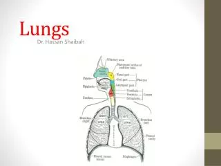
Lungs Digital Laboratory
Lungs Digital Laboratory. It’s best to view this in Slide Show mode, especially for the quizzes. This module will take approximately 75 minutes to complete . After completing this exercise, you should be able to:

Lungs Digital Laboratory
E N D
Presentation Transcript
Lungs Digital Laboratory It’s best to view this in Slide Show mode, especially for the quizzes. This module will take approximately 75 minutes to complete.
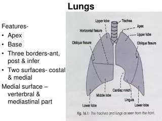
After completing this exercise, you should be able to: • Distinguish, at the light microscope level, each of the following organs and their specific features • Bronchus – respiratory epithelium, lamina propria, glands, hyaline cartilage, smooth muscle • (no need to differentiate between primary, secondary, or tertiary) • Bronchiole - respiratory epithelium, lamina propria, smooth muscle • (regular) bronchiole • Terminal bronchiole • Respiratory bronchiole • Alveolar ducts • Alveolar sacs • Alveoli • Pulmonary artery • Bronchial (bronchiolar) artery • Pulmonary vein • Visceral Pleura • Alveolar macrophage / Dust Cell • Distinguish, at the electron microscopic level, each of the following cells or structures • Type I pneumocytes (type I alveolar cells) • Type II pneumocytes (type II alveolar cells) • Lamellar bodies • Basal lamina • Endothelial cells • Components of connective tissue (fibroblast, elastic fibers, etc.) if present • Alveolar macrophage (Dust cell)
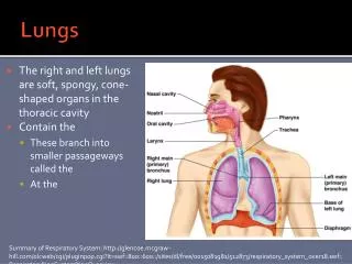
GROSS ANATOMY OF THE LUNG • Look and learn. Note: • Bronchi • Bronchioles • (regular) Bronchioles • Terminal bronchioles • Respiratory bronchioles • Alveoli • Alvoelar ducts • Alveolar sacs • As the respiratory passages decrease in diameter, changes include: • Breakup and then loss of cartilage • Decrease in number of glands • Relative increase in smooth muscle • Decrease in height of the epithelium
GROSS ANATOMY OF THE LUNG • Look and learn. Note: • Bronchi • Bronchioles • (regular) Bronchioles • Terminal bronchioles • Respiratory bronchioles • Alveoli • Alvoelar ducts • Alveolar sacs Bronchi have cartilage and glands, whereas bronchioles have neither of these. Bronchioles narrow in diameter as they get closer to the alveoli. The smallest bronchioles have some alveoli associated with them and are called respiratory bronchioles. The bronchioles immediately proximal to respiratory bronchioles are called terminal bronchioles, while the remaining bronchioles are simply called bronchioles. Alveolar ducts are long passages with alveoli, alveolar sacs are circular spaces leading to alveoli.
BRONCHUS Video showing proximal primary bronchus – slide 20 • Link to SL 020 • Be able to identify: • bronchus • Mucosa • Respiratory epithelium • Thick basement membrane • Lamina propria • Glands • Hyaline cartilage plates • Smooth muscle
BRONCHUS Video showing hilar primary bronchus – slide 24 • Link to SL 024 • Be able to identify: • bronchus • Mucosa • Respiratory epithelium • Thick basement membrane • Lamina propria • Glands • Hyaline cartilage plates • Smooth muscle
BRONCHI AND BRONCHIOLES blood vessels bronchus bronchus cartilage Bronchi have cartilage and glands, whereas bronchioles have neither of these. Since it’s sometimes difficult to see glands in small bronchi (image above), looking for cartilage is a more reliable way to differentiate bronchi from bronchioles. Also, note that both bronchi and bronchioles have a pseudostratified or columnar epithelium; these taller, thinner cells results in nuclei that are close together, creating a basophilic inner lining. Contrast this with blood vessels; the simple squamous epithelium of the tunica intima has nuclei that are spread out, so these are less basophilic near the lumen. We’ll deal with specific vessel identification shortly. bronchiole
BRONCHI AND BRONCHIOLES Video showing bronchi vs. bronchioles – slide 112 • Link to SL 112Aand SL 112Band SL 113and SL 059and SL 114 • Be able to identify: • bronchus • Mucosa • Respiratory epithelium • Thick basement membrane • Lamina propria • Glands • Hyaline cartilage plates • Smooth muscle • bronchiole • Mucosa • Respiratory epithelium • Thick basement membrane • Lamina propria • Smooth muscle
QUIZ Self-check: Identify the structure / organ from which these slides were taken. (advance slides for answers) ureter
VASCULATURE OF THE LUNG • Look and learn. Note: • Pulmonary artery • Bronchial artery • Pulmonary vein The pulmonary arteries are under low pressure, so it’s difficult to distinguish these from veins based on thickness of their walls. The bronchus/bronchiole, pulmonary artery, and bronchial artery are bundled together in the middle of a lobule. The pulmonary artery has the same approximate diameter as the bronchus or bronchiole that accompanies it. The bronchial artery is within the wall of , and much smaller in diameter than, the bronchus or bronchiole it supplies. The pulmonary veins are found in the partitions between the lobules, away from the bundled bronchus/bronchiole, pulmonary and bronchiole arteries.
VASCULATURE OF THE LUNG bronchiole bronchus The bronchus/bronchiole, (branch of the) pulmonary artery, and bronchial / bronchiolar artery are bundled together (orange outline) in the middle of a lobule. The pulmonary artery (red arrows) has the same approximate diameter as the bronchus or bronchiole that accompanies it. The bronchial / bronchiolar artery (blue arrows) is within the wall of , and much smaller in diameter than, the bronchus or bronchiole it supplies.
VASCULATURE OF THE LUNG The pulmonary veins (green arrow) are found in the partitions between the lobules, away from the bundled bronchus/bronchiole, pulmonary and bronchial / bronchiolar arteries. (red arrow is pulmonary artery) There are small lymphatic vessels associated with the bronchi and bronchioles, as well as in the partitions between lobules; these are difficult to definitively identify on our slides.
VASCULATURE OF THE LUNG Video showing pulmonary vasculature – slide 112 • Link to SL 112Aand SL 112Band SL 113and SL 059and SL 114 • Be able to identify: • Pulmonary arteries • Bronchial / bronchiolar arteries (arterioles) • Pulmonary veins
VASCULATURE OF THE LUNG FYI: The pulmonary arteries and most bronchial arteries drain into the capillaries supplying the alveoli, which then feed into the pulmonary veins. However, proximal bronchial arteries (i.e. those away from the alveoli) supply capillaries that feed into bronchial veins (see orange arrow in drawing, likely bronchial vein indicated in image to the right). You do not have to definitively identify bronchial veins.
TERMINAL AND RESPIRATORY BRONCHIOLES The transition from bronchioles to alveoli is gradual. Therefore, the smallest bronchioles have some alveoli associated with them and are called respiratory bronchioles. The bronchioles immediately proximal to respiratory bronchioles are called terminal bronchioles. X Here you can see respiratory bronchioles (green lines) partially lined with respiratory epithelium, and partially lined with alveoli. The terminal bronchiole (X) is connected to the respiratory bronchiole and is completely lined with respiratory epithelium.
TERMINAL AND RESPIRATORY BRONCHIOLES Video showing terminal and respiratory bronchioles – slide 114 • Link to SL 112Aand SL 112Band SL 113and SL 059and SL 114 • Be able to identify: • Terminal bronchioles • Respiratory bronchioles • Note these are difficult to find, and may require some interpretation (imagination).
ALVEOLI, ALVEOLAR DUCTS, ALVEOLAR SACS The alveoli are the terminal structures of the respiratory tract. They are lined with simple squamous epithelium, with connective tissue within their walls. alveolus Ignore the green arrows for now. alveolus alveolus alveolus
ALVEOLI, ALVEOLAR DUCTS, ALVEOLAR SACS The terminal portions of the respiratory tract that lead into individual alveoli can be classified based on shape as either alveolar ducts (between red arrows) or alveolar sacs. Alveolar sac
ALVEOLI, ALVEOLAR DUCTS, ALVEOLAR SACS Video showing alveolar ducts and sacs – slide 112 • Link to SL 112Aand SL 112Band SL 113and SL 059and SL 114 • Be able to identify: • Alveoli • Alveolar ducts • Alveolar sacs
ULTRASTRUCTURE OF ALVEOLI • In the drawing, you can see that the alveolar cells are of three types: • type I alveolar cells (type I pneumocytes) – simple squamous, labeled alveolar epithelial cell, part of blood-air barrier • type II alveolar cells (type II pneumocytes – cuboidal, labeled septal cell, produces surfactant • Alveolar macrophages (dust cells) • The walls of the alveoli contain loose connective tissue with lots of elastic fibers and capillaries.
ULTRASTRUCTURE OF ALVEOLI • Our slides are sectioned too thick to see much detail in the alveoli. However, this sweet image from Wheater’s text is a thin resin section which shows good cellular detail: • P1 – type I pneumocyte (squamous, lines alveoli) • P2 – type II pneumocyte (cuboidal, also part of alveolar lining) • M – alveolar macrophage • C – capillary • E – endothelial cell Scanning EM of similar cut – sweet!!!
ULTRASTRUCTURE OF ALVEOLI air air air Electron micrograph from the lung: Endothelial cell Fused basal lamina Type I pneumocyte Elastic tissue RBC in capillary Endothelial cell nucleus Fibroblast in inter-alveolar space
ULTRASTRUCTURE OF ALVEOLI • Electron micrograph from the lung detailing the blood-air barrier: • P1 – type I pneumocyte • E – endothelial cell • BM – fused basal lamina • Er - erythrocyte Endothelial cells usually have more caveoli/pinocytotic vesicles than type I pneumocytes, but not on this image. air
ULTRASTRUCTURE OF ALVEOLI Type II pneumocytes have lamellar bodies (orange arrows) and form tight junctions (red arrows) with type I cells (blue arrows). air blood
ULTRASTRUCTURE OF ALVEOLI Electron micrograph from the fetal lung: Capillary RBC in capillary WBC in capillary Glycogen Lamellar bodies in alveolar space Type II pneumocyte Lamellar bodies in type II pneumocyte Macrophage (dust cell) Type II pneumocytes have lamellar bodies and form tight junctions (red arrows) with type I cells (blue arrows). Alveolar macrophages also have lamellar bodies, but often in lysosomes showing different stages of digestion. Macrophages do not make tight junctions with type I or type II cells. This is a fetal lung; amniotic fluid in the alveoli prevent the lamellar bodies from unraveling upon release.
ALVEOLAR MACROPHAGES (DUST CELLS) Note that dust cells can be found in the tissue or lumen of alveoli. • Link to SL 113and SL 059 • Be able to identify: • Alveolar macrophages (dust cells) • We didn’t have you look at our slides for the cell types in alveoli. It might be useful to spend a minute looking now to appreciate the difficulty one would have to distinguish most alveolar cells on our slides.
VISCERAL PLEURA Video showing visceral pleura – slide 114 • Link to SL 113and SL 059and SL 114 • Be able to identify: • Visceral pleura • You also may want to look at SL 184, which is the elastic stain. The left tissue is lung – purple/black is elastic fibers
The next set of slides is a quiz for this module. You should review the structures covered in this module, and try to visualize each of these in light and electron micrographs. • Distinguish, at the light microscope level, each of the following organs and their specific features • Bronchus – respiratory epithelium, lamina propria, glands, hyaline cartilage, smooth muscle • (no need to differentiate between primary, secondary, or tertiary) • Bronchiole - respiratory epithelium, lamina propria, smooth muscle • (regular) bronchiole • Terminal bronchiole • Respiratory bronchiole • Alveolar ducts • Alveolar sacs • Alveoli • Pulmonary artery • Bronchial (bronchiolar) artery • Pulmonary vein • Visceral Pleura • Alveolar macrophage / Dust Cell • Distinguish, at the electron microscopic level, each of the following cells or structures • Type I pneumocytes (type I alveolar cells) • Type II pneumocytes (type II alveolar cells) • Lamellar bodies • Basal lamina • Endothelial cells • Components of connective tissue (fibroblast, elastic fibers, etc.) if present • Alveolar macrophage (Dust cell)
QUIZ Self-check: Identify the entire structure, and 1-5. (advance slides for answers)
QUIZ Self-check: Identify entire structure in middle, and 1-3, (and 4?) (advance slides for answers)
QUIZ Self-check: Identify 1-4. (advance slides for answers) Note, this is a section of a terminal bronchiole. You didn’t look at these specifically in this module, but maybe you can figure these out.
QUIZ Self-check: Identify 1-6. (advance slides for answers)
QUIZ Self-check: Identify 1-5. (advance slides for answers)
QUIZ Self-check: Identify 1-6. (advance slides for answers)
QUIZ Self-check: Identify these cells. (advance slides for answers) Type I pneumocyte Type II pneumocyte X
QUIZ Self-check: Identify three structures. (advance slides for answers) X Pulmonary artery Bronchiolar (bronchial) artery X bronchiole
QUIZ Self-check: Identify these structures. (advance slides for answers) Terminal bronchiole Respiratory bronchiole
QUIZ Self-check: Identify the structure and tissue. (advance slides for answers) Smooth muscle bronchiole X
QUIZ Self-check: Identify these cells and spaces. (advance slides for answers) Blood or air? Blood or air? air Blood or air? Type II pneumocyte X X Endothelial cell Blood or air? Blood or air?
QUIZ Self-check: Identify these cells. (advance slides for answers) Alveolar macrophages / dust cells
QUIZ Self-check: Identify cell, outlined structure, and structure between arrows. (advance slides for answers) Elastic fibers Fused basal lamina Type I pneumocyte
QUIZ Self-check: Identify this structure. (advance slides for answers) Pulmonary vein X
QUIZ Self-check: Identify these cells. (advance slides for answers) fibroblast Endothelial cell
QUIZ Self-check: Identify these three structures. (advance slides for answers) bronchiole X X Bronchiolar (bronchial) artery Pulmonary artery