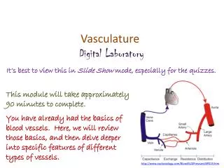
Vasculature Digital Laboratory
Vasculature Digital Laboratory. It’s best to view this in Slide Show mode, especially for the quizzes. This module will take approximately 90 minutes to complete .

Vasculature Digital Laboratory
E N D
Presentation Transcript
Vasculature Digital Laboratory It’s best to view this in Slide Show mode, especially for the quizzes. This module will take approximately 90 minutes to complete. You have already had the basics of blood vessels. Here, we will review those basics, and then delve deeper into specific features of different types of vessels. http://www.cvphysiology.com/Blood%20Pressure/BP019.htm
After completing this exercise, you should be able to: • Distinguish, at the light microscope level, each of the following:: • Structure of typical vessel • Tunics • Tunica intima • Tunica media • Tunica externa (adventitia) • Elastic laminae • Internal elastic laminae • External elastic laminae • Cell types • Fibroblasts • Smooth muscle cells • Endothelial cells • Arteries • Elastic (large) arteries • Muscular (medium) arteries • Arterioles • Veins • Venules • Medium veins • Large veins • Capillaries • Lymphatic channels • Distinguish, at the electron microscope level, each of the following:: • Capillaries • Continuous • Fenestrated • Discontinuous • Cells • Endothelial cells • Pericytes
Blood and lymphatic vessels Vessels in the body transport fluids, either blood or lymphatic fluid, which allows for distribution of nutrients, waste products, hormones, etc. throughout the body. There are two components of the vascular system: Cardiovascular system – transports blood, and includes the heart, arteries, capillaries, and veins Lymphatic system – which returns tissue fluid (lymph) from the tissues back to the cardiovascular system
Blood and lymphatic vessels Both of these systems are essentially a set of tubes, the walls of the tubes having different histological features reflecting their function. The contents of these tubes, namely blood and lymph, have been covered elsewhere. Here, we focus on the tubes themselves. This first section will provide the basic features of blood vessels, as well as differentiation of arteries and veins. Next, we will take a more in-depth look at the structure of a basic blood vessel. This will be followed by more detailed consideration of the different types of vessels.
Blood and lymphatic vessels Vessels have three basic components (from inside to outside): Tunica intima – a simple squamous epithelium, called the endothelium, with underlying loose connective tissue Tunica media – a thicker layer with smooth muscle and elastic fibers Tunica externa(adventitia) – dense connective tissue
Blood and lymphatic vessels In this cross section of a blood vessel, the blood in the lumen is indicated. The double-arrow indicates the extent of the tunica media; you should recognize the smooth muscle tissue that is predominant in this region. The smooth muscle cells are circularly arranged; in other words, the cells lie perpendicular to the long axis of the vessel. The thin portion of the wall “inside” the tunica media is the tunica intima, while the dense irregular connective tissue surrounding the tunica media is the adventitia. blood
Blood and lymphatic vessels In the higher-powered image, it is easier to see the tunica intima (green double-arrow), tunica media (black double-arrow) and adventitia (blue double arrow). Arrows indicate nuclei of endothelial cells that form the inner lining of vessels. blood The border between the intima and media (yellow dotted line) is indeed wavy here; we’ll address this in detail later.
Blood and lymphatic vessels Video of vessel layers – SL85 • Link to SL 085 • Be able to identify: • Blood vessel • Tunica intima • Tunica media • Tunica externa (adventitia)
Blood and lymphatic vessels • To begin distinguishing the major categories of vessels, note that: • The major histological difference between arteries and veins lies in the thickness and muscularity of the tunica media; arteries have a thicker, more muscular tunica media • Capillaries are composed simply of endothelial cells (and their basement membrane), without a tunica media or adventitia • Lymphatic vessels have an even less-developed tunica media, and the smallest lymphatic vessels have valves
Blood and lymphatic vessels Histologically, differentiating between arteries/arterioles (red arrow) and veins/venules(blue arrow) is best done by comparing vessels of approximately the same size. Arteries have a thicker smooth muscle layer in their wall; therefore, their wall is relatively thicker compared to the size of the vessel itself, with a narrower lumen. In addition, arteries tend to be rounder. Both will typically contain blood cells in their lumen, though during tissue preparation they can be washed away (see vessel toward the left) and become trapped in inappropriate locations. Arterioles are smaller arteries; venules are smaller veins.
Blood and lymphatic vessels Higher-powered view of the same vessels… All blood vessels are lined by a simple squamous epithelium, referred to as an endothelium. Endothelial cells have flattened nuclei (black arrows). The “fatter” nuclei in the wall of the vessel (green arrow), particularly in the artery, belong to smooth muscle cells.
Blood and lymphatic vessels Capillaries are very thin-walled, small vessels; they consist mostly of the endothelial cells. Because of this, they are difficult to find and identify definitively. Possible candidates for capillaries are indicated by the arrows, though the left is likely a small venule, and the right a small arteriole.
Blood and lymphatic vessels A couple more examples of capillaries. In the left image, note the simple squamous endothelial cells that make up the wall, and a diameter that is approximately the size of a red blood cell (possible RBC in upper capillary). To the right is a longitudinal section through a capillary (green arrows).
Blood and lymphatic vessels Lymphatic vessels (green arrows) typically have even thinner walls than veins, although these examples show relatively thick walls. However, small lymphatic vessels in tissues have numerous valves. In addition, they are typically devoid of red blood cells, and are filled with a “colloid” from precipitated lymph fluid as well as white blood cells. Yes, veins have valves too, but not in the smallest veins in the wall of an organ like we are looking at now. artery
Blood and lymphatic vessels Enlarged portion of previous image. If you look VERY closely in the artery, you might see that the pink disks are red blood cells, whereas the pink in the lymph vessels isn’t cellular. In addition, note the much greater proportion of white blood cells in the lymphatic vessels (green arrows) compared to the artery. artery
Blood and lymphatic vessels Video of lung hilus showing blood and lymphatic vessels – SL24 • Link to SL 024 • Be able to identify: • Artery / arteriole • Vein / venule • Capillary • Lymphatic channel
Detailed examination of blood vessel structure As mentioned above, vessel walls are made up of three layers: Tunica intima – forms the inner lining of the blood vessel, and includes the simple squamous endothelial cells, a basement membrane, and a subendothelial layer that is loose connective tissue. Tunica media – contains varying amounts of smooth muscle cells, elastic and collagen fibers Tunica externa(adventitia) – outer layer, typically dense irregular connective tissue, contains abundant collagen fibers secreted by fibroblasts
Detailed examination of blood vessel structure In the higher-powered image, it is easier to see the tunica intima (green double-arrow), tunica media (black double-arrow) and adventitia (blue double arrow). Arrows indicate nuclei of endothelial cells that form the inner lining of vessels. blood
Detailed examination of blood vessel structure The tunica intima has three components. As mentioned before, the brown arrows indicate endothelial cells, simple squamous cells that line the lumen. The loose connective tissue (X) below the endothelial cells is the subendothelial layer. The basement membrane between these two cannot be seen. X X
Detailed examination of blood vessel structure The tunica media in this vessel is predominantly smooth muscle cells, the nuclei of which are atypically long in this vessel (black arrows). The smooth muscle cells secrete most of the extracellular material (outlined), which includes elastic fibers, but also some collagen.
Detailed examination of blood vessel structure The tunica externa (adventitia) is dense irregular connective tissue, so consists of mostly collagen (green arrows), with some elastic fibers, both of which are secreted by the resident fibroblasts.
Detailed examination of blood vessel structure In many vessels, there are elastic sheets that separate the layers of the blood vessel wall: Internal elastic lamina – between the tunica intima and tunica media External elastic lamina – between the tunica media and tunica externa Some rules of thumb regarding the elastic laminae: They are more typical in arteries than veins They are more typical in larger vessels than smaller ones The external elastic lamina is not common but the internal elastic lamina can be seen in many vessels.
Detailed examination of blood vessel structure The elastic lamina are brightly eosinophilic bands that are interposed between the tunicas. In this vessel, the internal elastic lamina (purple arrows) is very apparent, while the external elastic lamina (blue arrows) is harder to see; nevertheless, its position can be deduced by recognition of the border between the media and externa.
Detailed examination of blood vessel structure Video of details of vessel wall – SL85 Video of details of vessel wall – SL115 • Link to SL 085and SL 115 • Be able to identify: • Tunica intima • Endothelial cells • (subendothelial layer) • Tunica media • Smooth muscle cells • Connective tissue • Tunica externa (adventitia) • Elastic lamina • Internal elastic lamina • External elastic lamina
Detailed examination of blood vessel structure The only term we didn’t define in this drawing is the vasa vasorum (it’s not in the objectives). This literally means “vessels of the vessels”; small blood vessels within the wall of larger vessels. As you know, diffusion only is efficient over small distances, so blood in the lumen of a vessel can only provide oxygen to the tunica intima and possibly the inner portion of the tunica media. However, the outer layers of a vessel require nutrients also. The vasa vasorum supply cells in the outer regions of the blood vessel, and are derived from branches of the vessel itself in the case of an artery (see red arrow), or from a nearby artery in the case of a vein.
TYPES of blood vessels Now that we have established the basic structure of a typical blood vessel, lets look at the different types of vessels. Arteries and veins can be broken down into three types each. As we will see, these distinctions are not based purely on size, but on histological features as well. In addition, there are three types of capillaries whose different histological features reflect functional differences between these vessels. This categorization is convenient. However, the transition between the different types of vessels is gradual, so many vessels will be “between” categories (i.e. a vessel may have characteristics of both an elastic and muscular artery). You will never see these “tweeners” on an exam.
TYPES of blood vessels Some of these vessel types have more than one name…these are indicated. aka - large artery aka - medium artery In case you care, I prefer to use the terms elastic and muscular arteries, since, as we will see, these terms describe the histology of these vessels more effectively.
TYPES of blood vessels The easiest thing to do instructionally is to start with vessels you have looked at previously. These are the vessels within the rectangle; the vessels we have been using as our “typical” arteries and veins. Since you have already seen these, there’s not much to say structurally. Texts differentiate muscular arteries and arterioles based on the number of layers of smooth muscle cells in the tunica media. (e.g. arterioles have 1 or 2 layers of smooth muscle, muscular arteries from 3-40 layers). Same for venules and medium veins. However, the numbers used by different texts vary, and some add “small arteries” as a category, so don’t get bent out of shape about counting layers of smooth muscles cells.
TYPES of blood vessels Physiologically, however, the difference between arterioles and muscular arteries is substantial. You will soon learn that the diameter of blood vessels is controlled largely by the state of contraction of the smooth muscle in the tunica media. Naturally, smaller vessels have both a smaller lumen, and less substantial total amount of smooth muscle. However, proportionally, arterioles have a higher smooth muscle / lumen ratio than muscular arteries, even if this distinction is subtle histologically. Therefore, contraction of smooth muscle in arterioles has a more significant impact on vessel diameter, making arterioles the vessels that have a larger impact on blood flow regulation. OK, enough of that silly-talk, back to histology…..
TYPES of blood vessels Lumen of muscular artery Tunica intima Tunica media Tunica externa Lumen of medium vein The large vessels we have been looking at in slide 85 are a muscular artery and a medium vein. The lumen of each vessel is indicated, as are the tunics (see key to left). Note that the tunica media of the muscular artery contains substantially more smooth muscle than the medium vein. The internal elastic lamina is also more readily visible in the artery. Traditionally, one considers the tunica externa of the vein to be more substantial than the artery, but this is not obvious on this slide.
TYPES of blood vessels Video of medium artery and vein– SL85 • Link to SL 085 • Be able to identify: • Muscular artery • Medium vein • (all regions and structures previously mentioned)
TYPES of blood vessels In this image, we see smaller versions of a medium vein (top) and muscular artery (bottom). Again, note that the artery has more smooth muscle in the tunica media, and a more prominent internal elastic lamina (bright eosinophilic band indicated by the black arrows). The artery is rounder due to smooth muscle (and elastic fibers) in the tunica media, while the vein has an irregular shape. Technically speaking, medium veins have valves, but you won’t see them on our slides (mostly because valves are in the more sizable medium veins).
TYPES of blood vessels Even smaller vein (blue outline) and artery (red). These are close to the size of venules and arterioles. The internal elastic lamina (black arrows) is less substantial in the artery/arteriole than in larger muscular arteries. Actually, that’s either two veins, or two profiles of the same vein as it turns.
TYPES of blood vessels Here you can see an arteriole (yellow outline) and venule (blue outline). Note the arteriole has a more muscular wall, and is rounder. The shape of the venule is irregular, and has less smooth muscle. Don’t forget that when identifying arteries from veins, or arterioles and venules, it’s best to choose pairs so that you can make a proper comparison.
TYPES of blood vessels Video of medium artery and vein to arterioles and venules – SL96 • Link to SL 096 • Be able to identify: • Muscular artery • Medium vein • Arteriole • Venule
TYPES of blood vessels In this electron micrograph of an arteriole, the internal elastic lamina is prominent (red arrows – recall that elastic tissue stains poorly in electron micrographs). Endothelial cell (A) and smooth muscle cell (B) nuclei are indicated. A B A Red blood cell
TYPES of blood vessels Another electron micrograph of an arteriole, in this case the internal elastic lamina is not prominent. Endothelial cell (A), smooth muscle cell (B), and fibroblast nuclei (C) are indicated. C A B
TYPES of blood vessels – Elastic arteries As you would guess, elastic arteries are vessels that contain abundant elastic tissue, in the form of tubes of elastin between the smooth muscle cells in the tunica media. Elastic arteries are vessels that receive blood from the ventricles; aorta, pulmonary trunk, proximal portions of the brachiocephalic, left common carotid, left subclavian arteries. Physiologically, elastic arteries serve two purposes: --they expand during ventricular systole (contraction), accepting the bolus of ejected blood --they recoil during diastole (ventricular relaxation), propelling blood forward So, although they do not “contract” to create a propulsive force, they do assist the ventricles in maintaining blood flow.
TYPES of blood vessels – Elastic arteries The tunica media in elastic arteries is dominated by elastic fibers (yellow arrows). Most of the nuclei you see in the media belong to smooth muscle cells, which are largely responsible for secreting the elastic fibers. Tunica intima Tunica media Tunica externa
TYPES of blood vessels – Elastic arteries Video of elastic artery – SL31 • Link to SL 031 • Be able to identify: • Elastic artery
TYPES of blood vessels – Elastic arteries Tunica intima Tunica media Tunica externa This tissue was stained for elastic fibers, which highlight the elastic fibers within this vessel nicely (yellow arrows in pseudomagnification inset of tunica media). The large dark bands (orange arrows) are artifacts caused by folding of the vessel during tissue preparation.
TYPES of blood vessels – Elastic arteries Video of elastic artery elastic stain – SL184 • Link to SL 184 • Be able to identify: • Elastic artery
TYPES of blood vessels – large veins Large veins have a unique characteristic relative to all other vessels. In addition to the circularly-arranged smooth muscle in the tunica media, they have numerous, longitudinally-oriented bundles of smooth muscle in the tunica externa(adventitia)(smooth muscle cells oriented in the direction of the black double-arrows). The purpose of this substantial amount of extra smooth muscle is unclear. It is possible that these muscle bundles are involved in contraction similar to gut peristalsis, assisting to move venous return to the atria under low pressures. So, in a cross section of a large vein, the smooth muscle in the tunica media is cut longitudinally, while the smooth muscle in the tunica adventitia is cut in cross section. In a longitudinal section of a large vein……
TYPES of blood vessels – large veins This is a cross-section of a large vein (NOT a longitudinal section); it is thin walled, so it has collapsed. Tunica intima Tunica media Tunica externa The predominant feature of large veins is the numerous bundles of smooth muscle (one bundle outlined) in the tunica externa. Note that, like all vessels except capillaries, this vessel still has circularly arranged smooth muscle in the tunica media.
TYPES of blood vessels – large veins Video of large vein – SL87 In slide 52 (adrenal gland), the large vein is here. • Link to SL 087 and SL 052 • Be able to identify: • Large vein
TYPES of blood vessels – capillaries Capillaries are the smallest vessels, and consist of only endothelial cells and basement membrane. Obviously, the thin wall of these vessels maximizes diffusion and other transport mechanisms for efficient nutrient and waste exchange between the blood and surrounding tissues. We have already looked at capillaries on glass slides (a still image is on the next slide so you don’t have to go back and find it). In this section, we want to focus on the different types of capillaries, which is based on their ultrastructure (electron micrographs).
Blood and lymphatic vessels Capillaries are very thin-walled, small vessels; they consist mostly of the endothelial cells. Because of this, they are difficult to find and identify definitively. Possible candidates for capillaries are indicated by the arrows, though the left is likely a small venule, and the right a small arteriole.
TYPES of blood vessels – capillaries The classification of capillaries is based on their ultrastructural characteristics, which relate to the permeability of the vessel. These include: Continuous capillaries – endothelial cells with tight junctions, complete basement membrane Fenestrated capillaries – endothelial cells with tight junctions, complete basement membrane, endothelial cells have fenestrations (pores) in them Discontinuous capillaries (aka sinusoids, not shown in drawing) – endothelial cells with fenestrations and gaps between the cells, incomplete basement membrane As I hope you deduced, these are listed in order of increasing permeability.
TYPES of blood vessels – capillaries Continuous capillaries consist of endothelial cells joined by tight junctions (cell junctions in image), and a complete basement membrane (basal lamina). Typically, endothelial cells in continuous capillaries have numerous pinocytotic vesicles. Pericytes form an incomplete layer of cells that share a basement membrane with endothelial cells. These cells have some contractile proteins, and may be active in phagocytosis. Continuous capillaries are the least permeable, and are located in tissues such as skeletal muscle, bone, cartilage, generic connective tissues.
TYPES of blood vessels – capillaries Fenestrated capillaries consist of endothelial cells joined by tight junctions and a complete basement membrane (basal lamina). The distinguishing feature is that the endothelial cells have numerous fenestrations (black arrows). In most places (except for the kidney), fenestrations contain a diaphragm (arrows in inset). Fenestrated capillaries typically have fewer pinocytotic vesicles than continuous capillaries. Fenestrated capillaries are more permeable than continuous capillaries, and are located in organs which require more robust exchange, or exchange of larger molecules, such as endocrine organs and some organs of the digestive tract.