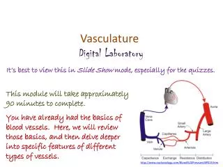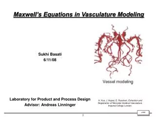
Vasculature
Vasculature. Spinal Cord. Brain. Anterior cerebral a. Anterior communicating a. Posterior communicating a. Middle cerebral a. Superior cerebellar a. Short C. A. Posterior cerebral a. Anterior inferior cerebellar a. Posterior inferior cerebellar a. Brain Stem. Brain Stem. PCA.

Vasculature
E N D
Presentation Transcript
Anterior cerebral a. Anterior communicating a. Posterior communicating a. Middle cerebral a. Superior cerebellar a. Short C. A. Posterior cerebral a. Anterior inferior cerebellar a. Posterior inferior cerebellar a.
Brain Stem PCA Sup. Cerebellar A. Pontine Branches Internal Auditory AICA Vertebral PICA
Anterior Choroidal • Frequently involved in strokes • Supplies • Optic system (optic tract, lat. Gen. nucl.) • Temporal nuclei (amygdala, hippocampus) • Internal capsule • GP and putamen • Thalamus
Cerebrum Anterior cerebral a. Anterior communicating a. Posterior communicating a. Middle cerebral a. Superior cerebellar a. Short C. A. Posterior cerebral a. Anterior inferior cerebellar a. Posterior inferior cerebellar a.
JN 6-6 JN 6-7 JN 6-7
Watershed Regionswhere to blood vessels run out distally, i.e., “border-zone regions with decreased blood supply
Venous sinuses In several locations the layers of dura separate to make venous dural sinuses. -Veins of the brain drain into a sinus --They have to “bridge” across the subarachnoid space and penetrate the arachnoid and dura to reach the sinus
Venous Sinuses S, superior sagittal sinus; L, lateral lacunae; G, arachnoid granulations; A, cerebral artery; C, cerebral vein; D, diploic vein; E, emissary vein; M, meningeal vein.
Venous sinuses Largest of the sinuses is: -Superior sagittal sinus Smaller ones; -inferior sagittal sinus: joins with the great cerebral vein to make the straight sinus -straight sinus runs in the tentorium and meets the superior sagittal sinus at the confluence of sinuses -transverse sinuses run laterally and become the sigmoid sinuses which become the internal jugular vein
Venous sinuses Superior sagittal sinus Inferior sagittal sinus Sigmoid sinus Great cerebral vein Straight sinus Transverse sinus Confluence of sinuses
Superior sagittal sinus Transverse sinus
Cavernous sinus Petrosal sinus Transverse sinus Sigmoid sinus Superior sagittal sinus