Download
1 / 37
400 likes | 901 Vues
Heart and Peripheral Vasculature. N1037. Anatomy and Physiology: Heart. Base @ top Apex @ bottom Pericardium Parietal layer Visceral layer. Anatomy and Physiology: Heart. 4 Chambers of the heart Right and left atria Right and left ventricles Heart valves Atrioventricular (AV) valves

E N D
Anatomy and Physiology: Heart • Base @ top • Apex @ bottom • Pericardium • Parietal layer • Visceral layer
Anatomy and Physiology: Heart • 4 Chambers of the heart • Right and left atria • Right and left ventricles • Heart valves • Atrioventricular (AV) valves • Tricuspid • Mitral (bicuspid) • Semilunar valves • Pulmonic • Aortic
Direction of Blood Flow • Inferior & Superior Vena Cava to Right Atrium (RA), then into Right Ventricle (RV) • Venous blood flows to Pulmonic Valve to Pulmonary Artery (unoxygenated) to the lungs. • Lungs oxygenate blood. Pulmonary veins (oxygenated) to Left Atrium (LA). • Into LA through Mitral Valve to Left Ventricle (LV) and ejected through Aortic Valve into Aorta. • Aorta delivers oxygenated blood to body.
Coronary Circulation • Left main coronary artery • Left circumflex artery • Left anterior descending artery • Right coronary artery
Cardiac Cycle • Systole • Isovolumic contraction • Early systole • Late systole • Diastole • Isovolumic relaxation phase • Early and mid-diastolic filling periods • Atrial systole (atrial kick)
Cardiac Cycle Diastole • Ventricles relax and fill with blood • The AV valves(tricuspid & mitral) are open • During the first rapid filling phase, blood pours rapidly from the atria into the ventricles (early diastolic filling). • At the end, the atria contract & push the last amount of blood into the ventricles (presystole = atrial kick). Systole • After this, the AV valves close and we hear the first heart sound “S1”. This is the beginning of Systole. • AV valves close to prevent regurgitation into the atria during contraction. • Then, the aortic and pulmonic valves (semilunar valves) open & blood is ejected rapidly into the arteries. • After all the contents are ejected, the semilunar valves close. This causes the second heart sound, “S2”. This is the end of systole.
Excitation of the Heart • Sinoatrial (SA) node • Atrioventricular node • Bundle of His • Right and left bundle branches • Purkinje fibers
Conduction Pathway and EKG • Sinoatrial node (SA Node) initiates an electrical impulse • It is the “pacemaker” of the heart. • Travels to the Atrioventricular node (AV Node) • Then it travels to the “Bundle of His” • Through the left and right bundle branches. • And lastly, through the ventricles.
Electrocardiogram (EKG) • P • Q • R • S • T • Isoelectric line
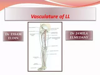
Peripheral Vasculature • Arterial system • Three layers of arterial walls: tunica intima, media, externa • Arteries • Arterioles • Capillaries • pulsating flow, no valves • Venous system • Veins • Venules • steady flow, 1 way valves,thinner walls , less elastic
Health History • Age • Childhood onset: rheumatic fever • Adult onset: HTN, CAD, MI, CVA, AAA • Gender • Female • Male • Race • May predispose to higher risk for CVA, CAD, HTN, diabetes mellitus
Common Chief Complaints • Chest pain • Syncope • Palpitations • Peripheral edema • Extremity pain Characteristics of Chief Complaints • Quality • Associated manifestations • Aggravating factors • Alleviating factors • Setting • Timing
Past Health History • Medical • Cardiac specific: AAA, angina, cardiogenic shock, chest trauma • Noncardiac specific • Surgical • Previous cardiovascular procedures • Common medications • Antianginals or vasodilators • Antidysrhythmics • Anticoagulants • Antihypertensives • Antilipemics • Diuretics • Inotropics • Thrombolytics
Past Health History • Communicable diseases • Childhood illnesses • Allergies • Aspirin • IVP dye • Seafood • Injuries and accidents
Family Health History • Assess for • Aneurysm • CVA • CAD • HTN • MI or sudden cardiac death • MVP
Social History • Alcohol, drug, or tobacco use • Sexual practices • Travel history • Work and home environment • Hobbies and leisure activities • Stress
Health Maintenance Activities • Sleep • Diet • Exercise • Stress management • Use of safety devices • Health check-ups
Risk Factors • Fixed • Age, gender, race, family history • Modifiable • HTN, hyperlipidemia, tobacco use, glucose intolerance, physical inactivity, diet, stress, sedentary lifestyle, obesity
Assessment Equipment • Equipment • Stethoscope • Sphygmomanometer • Watch with second hand • Tape measure
Inspection Ape To Man • Aortic 2ICS • Pulmonic 2ICS • Midprecordial 3ICS • Tricuspid 5ICS • Mitral 5ICS N = no visible pulsations except for the PMI in the mitral area
Palpation • Assess for pulsations, thrills, heaves • Assess the following areas: aortic, pulmonic, midprecordial, tricuspid, and mitral N = No pulsations, thrills, or heaves palpated, except in the mitral area, where the apical impulse may be palpated
Auscultation Use diaphragm and bell of stethoscope • N= Aortic: S2 is louder than S1 • N= Pulmonic: S2 is louder than S1 • N= Tricuspid: S1 is louder than S2 • N=Mitral: S1 is louder than S2 • Remember S1 = Apex, S2 = Base
Auscultation: Normal Findings • Aortic and Pulmonic • N= physiologoical split of S2 • Mitral and tricuspid: • N= S3 (gallop) may be heard in children, young adults, and pregnant women • N= S4 may indicate cardiac decompensation
Auscultation Abnormal • Murmurs • Use stethoscope diaphragm over aortic, pulmonic, mitral, and tricuspid areas • Use stethoscope bell over mitral and tricuspid areas • Possible causes • Characteristics: location, radiation, timing, intensity, quality, pitch, configuration • Pericardial friction rub • Characteristics: location, radiation, timing, quality, pitch • Possible cause
Assessment of Peripheral Vasculature • Inspection of jugular venous pressure • Place pt at 45°angle • measure vertical distance from sternum to top of distended neck vein • N= <4cm Abnormal > 4 cm indicates R ventricular pressure, bld vol, or obstruction
Inspection of Hepatojugular Reflux • Position pt at 30 ° in bed, press firmly on RUQ, observe neck for elevation of JVP N = no change in jugular veins Abnormal • A rise of more than 1 cm = right-sided CHF or fluid overload
Assessment of Arterial Pulses • Palpate temporal, carotid, brachial, radial, femoral, popliteal, posterior tibial, dorsalis pedis for rate, rhythm, amplitude, symmetry • auscultate with bell carotids, temporal & femoral pulses N= equal bilaterally, no bruits auscultated at carotids, temporal & femoral • Abnormal • Presence of bruits = obstruction due to atherosclerotic plaques, high-output states such as anemia or thyrotoxicosis
Special Techniques • Assessing for Pulsus paradoxus • take BP while pt supine,note 1st systolic sound heard, note point where all systolic sounds are not heard N= paradox should be < or = 10 mmHg • Abnormal • cardiac tamponade, pericardial effusion, cardiomyopathy, obstructive lung disease dt blood return to the L ventricle
Assessment of Peripheral Perfusion • Overall …..Evaluate peripheral pulses, color, clubbing, capillary refill, skin temperature, edema, ulcerations, hair distribution • Assess Venous system • Inspect fingers, legs, feet & toes • bend pts knee slightly and dorsiflex each foot - monitor for Homan sign N= no c/o calf pain Abnormal + VE Homan sign indicates DVT or thrombophlebitis
Assessment of Peripheral Perfusion • Assess Arterial system • Pallor test • Instruct pt to raise extremities • note the time it takes for pallor or lack of color to devlop N= no pallor develops within 60 secs • Allen test • ask pt to make fist , • occlude ulnar and radial artery • open hand and release one artery while compressing the other, repeat with opposite artery N= + ve Allen test = good blood flow both arteries in palm of hand Abnormal • no blood flow dt thrombus or atherosclerosis
Palpation of Epitrochlear Node • Place pt in supine position • support pt hand in your hand • plappate behind elbow in btwn biceps & triceps for epitrochlear node for size, shape, consistency , tenderness, & mobility N= node not palpable • Abnormal • enlarged lymph node
Gerontological Variations • Decreased size of heart muscle • Atria and ventricles become fibrotic and sclerotic • Decreased cardiac output • Change in heart position • Obesity • Vessels become fibrotic and rigid