Download
1 / 14
140 likes | 347 Vues
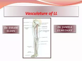
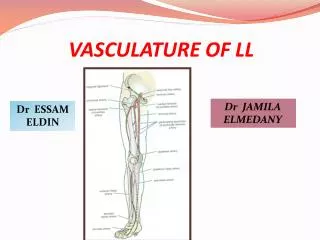
Vasculature Physiologic Disturbances. Blood Flow Studies Heart And Vessels. X-ray plain film for pulmonary vessels only Nuclear Medicine – V/Q Scan Angiography CT angiography Magnetic resonance angiography Ultrasound ( doppler ). Pulmonary Vascularity. Pulmonary arteries Pulmonary veins

E N D
Blood Flow StudiesHeart And Vessels • X-ray plain film for pulmonary vessels only • Nuclear Medicine – V/Q Scan • Angiography • CT angiography • Magnetic resonance angiography • Ultrasound (doppler)
Pulmonary Vascularity • Pulmonary arteries • Pulmonary veins • Bronchial arteries and veins • Interstitium • Lymphatics
Abnormal Pulmonic CirculationIncreased Pulmonary Circulation • Conditions causing increased pulmonary flow • Pulmonary hypertension. • Left-to-right shunt (VSD, ASD, PDA).
Abnormal Pulmonic CirculationIncreased Pulmonary Circulation • Pulmonary venous stasis and venous hypertension • Left heart dysfunction • Left heart failure, etc
Abnormal Pulmonic Circulation Decreased Pulmonary Circulation • Right ventricle outlet stenosis or obstruction • Pulmonary stenosis • Tetralogy of Fallot (VSD, Pulmonary stenosis, Overiding Aorta, RV hypertrophy) • Tricuspid valvular atresia, etc • Pulmonary artery stenosis or obstruction • Pulmonary infarct • Pulmonary embolism, etc
Pulmonary Emboli • PA chest • Essentially normal chest findings
Pulmonary Emboli • Lung scan • Ventilation scan with Xenon or particulate • Perfusion with multiple small emboli
Pulmonary Emboli • Multiple defects in the perfusion examination • Overall decreased perfusion in the right lung
Pulmonary Emboli • CT with contrast • Large low density filling defect in the right pulmonary artery • Exam of choice today
Pulmonary Emboli • Pulmonary angiogram • Catheter in left pulmonary artery from right femoral vein • Filling defect (thrombus) in the inferior interlobar branch of the left pulmonary artery
MRI Angiography • Non contrast technique possible • Visualize all of the vascular structures
Doppler Ultrasound • Doppler principles similar to that of the moving locomotive • Determine direction of flow and speed by the doppler (frequency) change of moving blood • Color assignment determined by direction of flow with respect to the transducer
Popliteal Vein Thrombus • Color doppler US • Axial image through popliteal • Flow in artery (red) • No flow in vein • Echogenic thrombus in vein (arrowhead)