Download
1 / 29
310 likes | 638 Vues
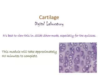
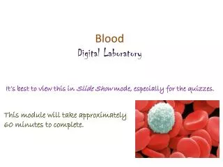
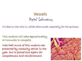
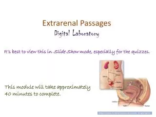
Extrarenal Passages Digital Laboratory. It’s best to view this in Slide Show mode, especially for the quizzes. This module will take approximately 40 minutes to complete . After completing this exercise, you should be able to:

E N D
Extrarenal Passages Digital Laboratory It’s best to view this in Slide Show mode, especially for the quizzes. This module will take approximately 40 minutes to complete.
After completing this exercise, you should be able to: • describe the basic three-layered structure of the extrarenal passages: mucosa, muscularis, adventitia • Calyxes • Ureter • urinary bladder • (female) urethra.
Organization of extra-renal passages • The major functions of the extra-renal passages include: • Transport and storage of urine • Maintenance of the ionic composition of the urine produced by the kidneys • To achieve these goals, the extra-renal passages have the following basic structure: • A mucosa, which consists of • transitional epithelium • lamina propria – an irregular connective tissue • A muscularis layer - composed of smooth muscle • An adventitia - composed of irregular connective tissue The qualities of each layer will vary somewhat depending on function. For example, the bladder will have a thick muscularis layer for urination (micturition). • The transitionial epithelium: • can stretch to allow filling (bladder) • is resistant to osmotic movement of ions and water, maintaining the concentration of urine produced by the kidneys urine
Minor calyx Urine passes out of the collecting ducts (papillary ducts) and into the minor calyces, and from there flows through the major calyces into the renal pelvis.
Minor calyx In these images, you can see a minor calyx (labeled PCS on the Wheater’s image) The major calyces and renal pelvis have histological features similar to the minor calyx, but without the papilla.
Minor calyx Video of Minor Calyx – SL115 • Link to SL 115 • Be able to identify: • Minor calyx • Transitional epithelium
ureter The three-layered histology of the extra-renal passages (mucosa, muscularis, adventitia) is more apparent in the ureter and bladder.
ureter Video of Ureter – SL18 • Link to SL 018 • Be able to identify: • ureter • mucosa • transitional epithelium • lamina propria • muscularis • adventitia
Urinary bladder The urinary bladder also has the characteristic three-layered histology of the extra-renal passages (mucosa, muscularis, adventitia). Here, the muscularis layer is quite thick to propel urine during micturition, and has three layers of smooth muscle.
Urinary bladder Video of Urinary Bladder – SL179 • Link to SL 179 • Be able to identify: • Urinary bladder • mucosa • transitional epithelium • lamina propria • muscularis • adventitia
Urinary bladder Video of Urinary Bladder (baby) – SL57 • Link to SL 057 • Be able to identify: • Urinary bladder • mucosa • transitional epithelium • lamina propria • muscularis • adventitia
bladder A final, minor note on the bladder: The abdominal cavity is lined with an epithelium, called peritoneum, that lines the outer surface of the abdominal organs (visceral peritoneum, red arrow), and the inner surface of the abdominal wall (parietal peritoneum, yellow arrow). This should be review from when you studied the gastrointestinal tract. Because of it’s position in the pelvic cavity, only a portion of the bladder is covered by peritoneum. Sections through most of the bladder (e.g. green line) do not include this peritoneal epithelium. In these cases, the outer layer of the bladder is simply connective tissue, and we call this outer layer an adventitia. However, a section near the apex of the bladder (blue line in image) will have an outer layer composed of connective tissue covered with parietal epithelium – we call this layer a serosa. Again, this is similar to what you learned in the GI section. The scanned digital slides have an adventitia as an outer layer. Some glass slides have a serosa.
urethra • The histological features of the urethra are different from other extra-renal structures: • The epithelium of the urethra near the bladder is transitional, but most of the urethra is lined by stratified squamous non-keratinized epithelium. • Small mucous glands may be present, but typically are not obvious on our slides. • The lamina propria has an abundant venous plexus • The smooth muscle is less developed, and may be interspersed with skeletal muscle of the pelvic floor (e.g. urogenital diaphram)
Urethra This section of the female urethra also includes the anterior wall of the vagina (see blue line in image). Therefore, you will see a cross section through the urethra - the lower portion of the slide, including the epithelium at the bottom, will be from the anterior wall of the vagina.
Urethra (female) Video of Urethra – SL120 • Link to SL 120 • Be able to identify: • Urethra • mucosa • Epithelium (transitional or stratified squamous) • lamina propria • muscularis • adventitia
The next set of slides is a quiz for this module. You should review the structures covered in this module, and try to visualize each of these in light and electron micrographs. • describe the basic three-layered structure of the extrarenal passages: mucosa, muscularis, adventitia • Calyxes • Ureter • urinary bladder • (female) urethra.
quiz Self-check: Identify structures 1-6. (advance slide for answers)
quiz Self-check answers
quiz Self-check: Identify the entire structure, and structures 1-6. (advance slide for answers)
quiz Self-check answers
quiz Self-check: Identify the entire structure, and structures 1-6. (advance slide for answers)
quiz Self-check answers Note: the epithelium of the urethra varies considerably
quiz Self-check: Identify the entire structure. bladder
quiz Self-check: Identify the entire structure. bladder
quiz Self-check: Identify the entire structure. urethra
quiz Self-check: Identify the entire structure. ureter Note that, like the bladder, the ureter can also have substantial adipose in the adventitia.
quiz Self-check: Identify the entire structure from which these images were taken. bladder
quiz Self-check: Identify the entire structure. ureter
quiz Self-check: Identify the structure indicated by the arrows. Minor calyx