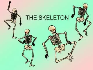
The Skeleton Part D
The Skeleton Part D. 7. Appendicular Skeleton. The appendicular skeleton is made up of the bones of the limbs and their girdles Pectoral girdles attach the upper limbs to the body trunk Pelvic girdle secures the lower limbs. Pectoral Girdles (Shoulder Girdles).


The Skeleton Part D
E N D
Presentation Transcript
The Skeleton Part D 7
Appendicular Skeleton • The appendicular skeleton is made up of the bones of the limbs and their girdles • Pectoral girdles attach the upper limbs to the body trunk • Pelvic girdle secures the lower limbs
Pectoral Girdles (Shoulder Girdles) • The pectoral girdles consist of the anterior clavicles and the posterior scapulae • They attach the upper limbs to the axial skeleton in a manner that allows for maximum movement • They provide attachment points for muscles that move the upper limbs
Pectoral Girdles (Shoulder Girdles) Figure 7.22a
Clavicles (Collarbones) • The clavicles are slender, doubly curved long bones lying across the superior thorax • The acromial (lateral) end articulates with the scapula, and the sternal (medial) end articulates with the sternum • They provide attachment points for numerous muscles, and act as braces to hold the scapulae and arms out laterally away from the body
Clavicles (Collarbones) Figure 7.22b, c
Scapulae (Shoulder Blades) • The scapulae are triangular, flat bones lying on the dorsal surface of the rib cage, between the second and seventh ribs • Scapulae have three borders and three angles • Major markings include the suprascapular notch, the supraspinous and infraspinous fossae, the spine, the acromion, and the coracoid process
Scapulae (Shoulder Blades) Figure 7.22d, e
The Upper Limb • The upper limb consists of the arm (brachium), forearm (antebrachium), and hand • Thirty-seven bones form the skeletal framework of each upper limb
Arm • The humerus is the sole bone of the arm • It articulates with the scapula at the shoulder, and the radius and ulna at the elbow
Arm • Major markings • Proximal humerus includes the head, anatomical and surgical necks, greater and lesser tubercles, and the intertubercular groove • Distal humerus includes the capitulum, trochlea, medial and lateral epicondyles, and the coronoid and olecranon fossae • Medial portion includes the radial groove and the deltoid process
Humerus of the Arm Figure 7.23
Forearm • The bones of the forearm are the radius and ulna • They articulate proximally with the humerus and distally with the wrist bones • They also articulate with each other proximally and distally at small radioulnar joints
Ulna • The ulna lies medially in the forearm and is slightly longer than the radius • Forms the major portion of the elbow joint with the humerus • Its major markings include the olecranon, coronoid process, trochlear notch, radial notch, and the styloid process
Radius • The radius lies opposite (lateral to) the ulna and is thin at its proximal end, widened distally • The superior surface of the head articulates with the capitulum of the humerus • Medially, the head articulates with the radial notch of the ulna • Major markings include the radial tuberosity, ulnar notch, and styloid process
Radius and Ulna Figure 7.24
Carpus (Wrist) • Consists of eight bones • Scaphoid, lunate, triquetral, and pisiform • Trapezium, trapezoid, capitate, and hamate • Some lovers try positions that they can handle
Metacarpus (Palm) • Five numbered (1-5) metacarpal bones radiate from the wrist to form the palm
Phalanges (Fingers) • Each hand contains 14 miniature long bones called phalanges • Fingers (digits) are numbered 1-5, beginning with the thumb (pollex) • Each finger (except the thumb) has three phalanges – distal, middle, and proximal • The thumb has no middle phalanx
Hand Figure 7.26a
Pelvic Girdle (Hip) • The hip is formed by a pair of hip bones (os coxae, or coxal) • Together with the sacrum and the coccyx, these bones form the bony pelvis • The pelvis • Attaches the lower limbs to the axial skeleton with the strongest ligaments of the body • Transmits weight of the upper body to the lower limbs • Supports the visceral organs of the pelvis
Pelvic Girdle (Hip) Figure 7.27a
Ilium • The ilium is a large flaring bone that forms the superior region of the coxal bone • It consists of a body and a superior winglike portion called the ala • Major markings include the iliac crests, four spines, greater sciatic notch, iliac fossa, and the pelvic brim
Ischium • The ischium forms the posteroinferior part of the hip bone • The thick body articulates with the ilium, and the thinner ramus articulates with the pubis • Major markings include the ischial spine, lesser sciatic notch, and the ischial tuberosity
Pubis • The pubic bone forms the anterior portion of the hip bone • It articulates with the ischium and the ilium • Major markings include superior and inferior rami, the pubic crestpubic symphysis, and obturator foramen (along with ilium and ischium)
Pubis: Lateral View Figure 7.27b
Pubis: Medial View Figure 7.27c
Comparison of Male and Female Pelvic Structure • Female pelvis • Tilted forward, adapted for childbearing • True pelvis defines birth canal • Cavity of the true pelvis is broad, shallow, and has greater capacity
Comparison of Male and Female Pelvic Structure • Male pelvis • Tilted less forward • Adapted for support of heavier male build and stronger muscles • Cavity of true pelvis is narrow and deep
Comparison of Male and Female Pelvic Structure Image from Table 7.4
The Lower Limb • The three segments of the lower limb are the thigh, leg, and foot • They carry the weight of the erect body, and are subjected to exceptional forces when one jumps or runs
Femur • The sole bone of the thigh is the femur, the largest and strongest bone in the body • It articulates proximally with the hip and distally with the tibia and fibula • Major markings include the head, greater and lesser trochanters, lateral and medial condyles and epicondyles, linea aspera, patellar surface, and the intercondylar notch
Femur Figure 7.28b
Leg • The tibia and fibula form the skeleton of the leg • They articulate with the femur proximally and with the ankle bones distally
Tibia • Receives the weight of the body from the femur and transmits it to the foot • Major markings include medial and lateral condyles, intercondylar eminence, the tibial tuberosity, anterior crest, medial malleolus
Tibia and Fibula Figure 7.29
Fibula • Sticklike bone with slightly expanded ends located laterally to the tibia • Major markings include the head and lateral malleolus
Foot • The skeleton of the foot includes the tarsus, metatarsus, and the phalanges (toes) • The foot supports body weight and acts as a lever to propel the body forward in walking and running Figure 7.31a
Tarsus • Composed of seven bones that form the posterior half of the foot • Body weight is carried primarily on the talus and calcaneus • Talus articulates with the tibia and fibula superiorly, and the calcaneus inferiorly • Other tarsus bones known as cuneiforms collectively
Tarsus Figure 7.31b, c
Calcaneus • Forms the heel of the foot • Carries the talus on its superior surface • Point of attachment for the calcaneal (Achilles) tendon of the calf muscles
Metatarsus and Phalanges • Metatarsals • Five (1-5) long bones that articulate with the proximal phalanges • Phalanges • bones of the toes • Each digit has three phalanges except the big toe, which has no middle phalanx
Metatarsus and Phalanges Figure 7.31a
Arches of the Foot • The foot has three arches maintained by interlocking foot bones and strong ligaments • Arches allow the foot to hold up weight • The arches are: • Lateral longitudinal – cuboid is keystone of this arch • Medial longitudinal – talus is keystone of this arch • Transverse – runs obliquely from one side of the foot to the other
Developmental Aspects: Fetal Skull • At birth, fetal skull bones are incomplete and connected by fontanels • Fontanels • Unossified remnants of fibrous membranes between fetal skull bones • The four fontanels are anterior, posterior, mastoid, and sphenoid
Developmental Aspects: Fetal Skull • Skull bones such as the mandible and maxilla are unfused Figure 7.33