Inflammatory Disorders
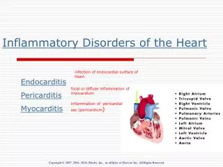
Inflammatory Disorders. Updated Spring 2012 by Nancy Jenkins. Layers of the Heart Muscle. TISSUES SURROUNDING THE HEART. Infective Endocarditis. Infection of the inner layer of the heart Usually affects the cardiac valves Was almost always fatal until development of penicillin


Inflammatory Disorders
E N D
Presentation Transcript
Inflammatory Disorders Updated Spring 2012 by Nancy Jenkins
Infective Endocarditis • Infection of the inner layer of the heart • Usually affects the cardiac valves • Was almost always fatal until • development of penicillin • Around 15,000 cases diagnosed • annually in the U.S.
Causative Organisms • Causative organism more virulent • Streptococcus viridans • Staphylococcus aureus • Viruses • Fungi
Etiology and Pathophysiology • Occurs when blood turbulence within heart allows causative agent to infect previously damaged valves (congenital or acqired valvular disease) or other endothelial surfaces • May occur in people with a history of rheumatic heart disease • May occur in people with normal valves with increased amounts of bacteria
Etiology/Pathophysiology • Endocarditis • When valve damaged, blood is slowed down and forms a clot. • Bacteria get into blood stream • Bacterial or fungal vegetative growths deposit on normal or abnormal heart valves • Vegetation • Fibrin, leukocytes, platelets, and microbes • Adhere to the valve or endocardium • Embolization of portions of vegetation into circulation
Classifications of Endocarditis • Acute Infective Endocarditis • Abrupt onset • Rapid course • Staph Aureus • Subacute Infective Endocarditis SBE- most common • Gradual onset • Systemic manifestations • Prosthetic Valve Endocarditis
Risk Factors- endocarditis • Hx of rheumatic fever or damaged heart valve • Prior history of endocarditis • Invasive procedures- (introduce bacteria into blood stream) (surgery, dental, etc) • Recent Dental Surgery • Permanent Central Venous Access • IV drug users • Valve replacements
Nursing Assessment • Subjective Data • History of valvular, congenital, or syphilitic cardiac disease • Previous endocarditis • Staph or strep infection • Immunosuppressive therapy-(HIV) • Recent surgeries and procedures
Nursing Assessment • Functional health patterns • IV drug abuse • Alcohol abuse • Weight changes • Chills
Nursing Assessment • Diaphoresis • Fever • Bloody urine • Exercise intolerance • Generalized weakness • Fatigue • Cough
Nursing Assessment • Dyspnea on exertion • Night sweats • Chest, back, abdominal pain
Collaborative Care • Fungal and prosthetic valve endocarditis • Responds poorly to antibiotics • Valve replacement is adjunct procedure
Assesment endocarditis • Infection and emboli • Emboli-spleen most often affected (splenectomy) • Osler’s nodes- painful, red or purple pea-sized lesions on toes and fingertips • Splinter hemorrhages- black longitudinal streaks on nail beds • Janeway lesions- flat, painless, small, red spots on palms and soles • Roth spots- hemorrhagic retinal lesions • Murmur- 90% have murmurs Heart Sounds Assessment Video • T above 101(blood cultures) and low-grade • Chills • Anorexia • Fatigue
Splinter hemorrhage • small areas of bleeding under the fingernails or toenails. • due to damage to capillaries by small clots
Janeway Lesions • flat, painless red spots on palms and soles
Osler’s Nodes • Painful, pea-size, red or purple lesions • On finger tips or toes Roth spots Osler’s nodes
Roth’s Spots • hemorrhagic retinal lesions
Diagnostic Tests • Blood Cultures- • Echocardiogram-TEE best- see vegetations • Other- WBC with differential, CBC,ESR, serum creatinine,CXR, and EKG
Echocardiogram-video • Mitral Valve involvement
Diagnostic Criteria • Duke criteria • 2 major • Blood cultures and vegetations present • 5 minor • symptoms
Treatment- Medications • Antibiotics • IV for 2-8 weeks • Monitor peaks and troughs of certain drugs • Monitor BUN and Creat. • Unclear success of oral antibiotics if not a good candidate for IV. Oral antibiotics are considered when dealing with endocarditis: • Of the tricuspid valve • With a causative organism sensitive to oral agents • Long-term IV therapy difficult or impossible • Outpatient f/u can be arranged
Nursing Diagnoses • Risk for Imbalanced Body Temperature • Risk for Ineffective Tissue Perfusion-emboli • Ineffective Health Maintenance
Complications • Emboli (50% incidence) • Right side- pulmonary emboli (esp. with IV drug abuse- Why??) • Left side-brain, spleen, heart, limbs,etc • CHF- 80% incidence with involvement of aortic valve • check edema, crackles, VS • Arrhythmias- A-fib • Death .
Prevention • Eliminate risk factors • Patient teaching
Risk Stratisfication for IE High Risk- • Mechanical prosthetic heart valve • Natural prosthetic heart valve • Prior infective endocardititis • Valve repair with prosthetic material • Most congenital heart diseases Moderate Risk- • Valve repair without prosthetic material • Hypertrophic cardiomyopathy • Mitral valve prolapse with regurgitation • Acquired valvular dysfunction Low Risk- • Innocent heart murmurs • Mitral valve prolapse without regurgitation • Coronary artery disease • People with pacemakers/ defibrillators • Prophylactic antibiotics are generally recommended only for people in the “High Risk” category
Collaborative Care • Prophylactic treatment for patients having • Removal or drainage of infected tissue • Renal dialysis • Ventriculoatrial shunts • Dental, oral, or upper respiratory tract procedures • Endocarditis video review
Myocarditis Myocarditis is an uncommon inflammation of the heart muscle (myocardium). This inflammation can be caused by infectious agents, toxins, drugs or for unknown reasons. It may be localized to one area of the heart, or it may affect the entire heart. (effects like pounding the heart get inflammation and swelling)
Etiology/Pathophysiology • Myocarditis-video • Virus, toxin or autoimmune response causes necrosis of the myocardium • Most often caused by viral infection • Frequently caused by Coxsackie B virus • Frequently follows an upper respiratory infection or viral illness • Get decreased contractility • Can become chronic and lead to dilated cardiomyopathy- heart transplant or death
This is an infection in the muscles of the heart, most commonly caused by the Coxsackie B virus that follows upon a respiratory or viral illness, bacteria and other infectious agents.
Risk factor-myocarditis • Hx of upper respiratory infection • Toxic or chemical effects (radiation, alcohol) • Autoimmune or immunosuppresents- 10% HIV develop it • Metabolic-lupus • Heat stroke or hypothermia
Assessment myocarditis • Infection and CHF • Fatigue,DOE • Tachycardia • Arrhythmias- PVCs, PACs, Atrial Tachycardias, • Chest pain • Signs of heart failure (S3, etc.) • Pericarditis frequently occurs with myocarditis- check friction rub
Diagnostic Tests • EKG- Non-specific T-wave abnormalities • CK-MB and Troponin may be elevated • Endomyocardial biopsy- there are risks and not used for every case but is definitive for myocarditis • Chest X-Ray- Variable (Normal to Cardiomegaly) • Echocardiogram • Cardiovascular Magnetic Resonace • A safe and sensitive noninvasive diagnostic test to confirm the diagnosis is not available
Medications • Antibiotics • Antiviral with interferon-a • IVIG- experimental trials • Corticosteroids or immunosuppressents • HF drugs- ACE, diuretics, beta blockers etc • Antiarrhythmics • Anticoagulants- Why??
Other Treatments • Bedrest and activity restrictions- Why important?? • **Activities may be limited for 6 months- 1 yr. • O2 GOAL- Decrease workload of the heart so it can heal
Nursing Diagnoses • Activity Intolerance • Decreased CO • Anxiety • Excess fluid Volume
Pericarditis • Pericarditis is an inflammation of the pericardium, the thin, fluid-filled sac surrounding the heart. It can cause severe chest pain (especially upon taking a deep breath) and shortness of breath.