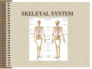
Overview of Skeletal System Functions and Bone Structure
520 likes | 622 Vues
Learn about the functions of the skeletal system, bone composition, classifications, bone development, repair of fractures, and skeletal divisions. Explore topics such as ossification, bone remodeling, types of fractures, and the role of bones as levers in movement.

Overview of Skeletal System Functions and Bone Structure
E N D
Presentation Transcript
SKELETAL SYSTEM FUNCTIONS • Support (Primary function) • Movement (Passive) • Protection of Vital Organs • Mineral Storage • Blood Cell Formation (Hematopoiesis or Hemopoiesis)
OSSEOUS C.T. • Compact (dense) Bone • Hard & heavy • Forms surface & diaphysis • Osteons = building blocks • Cancellous (spongy) Bone • Lightweight • Fills epiphyses, Contains red marrow • Trabeculae = building blocks • Matrix • Mineral Salts (hardness) • Collagen (strong & flexible)
Bone Cells • Osteoblasts – Secrete to form bone • Osteocytes • Mature bone cells • “Trapped” osteoblasts • Osteoclasts – destroy bone • Enzymes digest protein • Acids dissolve minerals • Forms Marrow Cavity; Involved in Remodeling
SKELETAL DIVISIONS • Axial • Appendicular
Classification: Shape & Location • Long • Short • Flat • Irregular • Sesamoid (develop in tendons; patella) • Sutural (between cranial bones)
LONG BONE ANATOMY • Diaphysis = shaft made of compact bone • Epiphyses = ends filled with spongy bone containing red marrow • Articular cartilage covers epiphyses • Epiphyseal line indicates earlier location of epiphyseal (growth) plate
LONG BONE ANATOMY • Periosteum is C.T. covering bone • Nutrient Foramina – holes allowing for penetration of arteries • Medullary cavity contains yellow marrow • Endosteum is C.T. lining medullary cavity
BONE DEVELOPMENT • Ossification = replacement of other connective tissue with bone • Begins during the 2nd month of gestation • Size increases until late teens (females) to mid-twenties (males) • Ossification processes include: • Intramembranous bone formation • Endochondral bone formation
INTRAMEMBRANOUS OSSIFICATION • Occurs in flat bones of skull, clavicles, mandible • Begins with fibrous C.T. membrane • Membrane calcifies & ossifies into bone • Fontanels • “Soft spot”, not yet ossified • Allows for birth & brain growth
Process of Intramembranous Ossification • Embryonic C.T. cells cluster & differentiate • Osteoblasts form & produce matrix = ossification center • Newly formed matrix calcifies • Osteocytes form
Process of Intramembranous Ossification • Trabeculae form; periosteum forms from surrounding condensed embryonic C.T. • Surface trabeculae fill with matrix, forming compact bone
ENDOCHONDRAL OSSIFICATION • Occurs in remainder of skeleton • Begins with hyaline cartilage model • Cartilage is replaced by bony tissue • Steps include: • Bone collar forms • Cartilage in shaft calcifies • Primary Ossification center forms in shaft • Secondary Ossification centers in epiphyses
APPOSITIONAL GROWTH • Bone Widens • Osteoclasts enlarge medullary cavity • Osteoblasts add bone to surface of diaphysis
BONE REMODELING • Replacement of old bone with new bone • Involves resorption (osteoclasts) & deposition (osteoblasts) • Alters bone shape in response to stress • Benefits: • Allows for growth • Removes injured bone • New bone is more resistant to fracture
FRACTURES AND THEIR REPAIR • Definition: Any break in a bone • Repair may take months • Requires: • Adequate minerals • Vitamins • Hormones • Weight-bearing exercise
STEPS IN FRACTURE REPAIR • Broken blood vessels form a hematoma (blood clot) • C.T. and Capillaries invade site; C.T. cells form fibrocartilage callus • Bony callus of spongy bone replaces fibrocartilage callus • Bony callus is remodeled
Types of Fractures • Open (Compound) – Broken bone ends protrude through the skin • Closed (Simple) – Bone does not penetrate the skin
Types of Fractures • Comminuted – splintered, crushed; small pieces between broken ends. Elderly. Most difficult to treat. • Greenstick – Partial fracture; one side breaks, other side bends. Children.
Types of Fractures • Impacted – One end of fractured bone forcefully driven into other end • Spiral – fracture spirals around long bone axis from twisting force
Types of Fractures • Pott’s – distal end of fibula, tibia or both • Colles’ – distal end of radius
Types of Fractures • Stress Fracture • Fracture without visible break • Microscopic fissures • No apparent damage to surrounding tissues • Result from repeated, strenuous activities • Can result from reduced calcium deposition in disease • 25% involve tibia
BONES AS LEVERS • Lever: A rigid rod that moves about a fixed point • Fulcrum: The fixed point around which a lever moves (joints) • Forces: Act to move levers at two points • Resistance: Force to be overcome • Effort or Work: Force required to overcome resistance; supplied by skeletal muscles
CLASSES OF LEVERS • First Class: The fulcrum is between the effort/force and the resistance • Seesaw • Tilting head backward
R R R R R R R F E E E E E E E FIRST CLASS LEVER
CLASSES OF LEVERS CONTINUED • Second Class: Resistance is between the fulcrum and the effort/force • Wheelbarrow • Rising up on one’s toes
R R R R R R R R F E E E E E E E E SECOND CLASS LEVER
CLASSES OF LEVERS CONTINUED • Third Class: The effort/force is between the fulcrum and the resistance • Most common type in the human body • Flexing the elbow
R R R R R R R R F E E E E E E E E THIRD CLASS LEVER
CLASSIFICATION OF JOINTS • Structural • Based on what tissues or structures are found between the bones • Fibrous, Cartilagenous, Synovial • Functional • Based on amount of movement (& amount of movement is determined by structures found between bones) • Freely movable, Slightly movable, Immovable
Pubic symphysis Functional: AmphiarthrosisStructural: Cartilagenous Knee Functional: DiarthrosisStructural: Synovial Sutures Functional: SynarthrosisStructural: Fibrous ARTICULATIONS: EXAMPLES
STRUCTURE OF A SYNOVIAL JOINT • Articular cartilage – covers bone ends • Synovial membrane – lines joint capsule • Synovial fluid – lubricates & nourishes cartilage • Synovial cavity – space between the bones • Joint capsule – fibrous C.T. • Ligaments – reinforce joint • Bursae – synovial sacs at other sites of friction


