Download
1 / 34
410 likes | 1.26k Vues
Neuroendocrine Responses to Stress. It is clear that endocrine responses constitute an integral component of the stress response.

E N D
It is clear that endocrine responses constitute an integral component of the stress response. Stress can affect the hormonal control of metabolism, reproduction, growth and immunity. Since hormone signalling plays a vital role in the maintenance of homeostasis, virtually every endocrine system responds in some fashion to specific stressors. The overall effect on the animal’s adaptive response to stress is an integration of multiple, and often interactive, hormone responses that directly affect physical health and well-being.
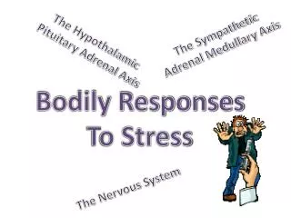
Neuroendocrinology can be defined as the study of communication between the central nervous system and endocrine glands. The term ‘neuroendocrine’ classically refers to hormone signalling involving the hypothalamus, pituitary gland and peripheral body systems. The hypothalamus is a bilaterally symmetric region on the inferior aspect of the brain above the anterior pituitary gland.
The pituitary gland comprises three components: (i) the anterior lobe; (ii) the posterior lobe; and (iii) the intermediate lobe. The anterior pituitary gland contains specialized cells that produce and secrete growth hormone, adrenocorticotropic hormone (ACTH), thyroid-stimulating hormone (TSH), luteinizing hormone (LH), folliclestimulating hormone (FSH), and prolactin. The chemical structures and major functions of the anterior pituitary hormones have been described elsewhere.
The role of the intermediate lobe is species specific, and in the human as well as a small number of other mammals the intermediate lobe is considered rudimentary. The posterior lobe of the pituitary is an extension of the floor of the third ventricle and maintains physical contact with the hypothalamus via the pituitary stalk. The posterior lobe acts as a storage site for vasopressin and oxytocin. Vasopressin, also known as antidiuretic hormone (ADH), enhances water reabsorption by the kidney, constricts smooth muscles surrounding blood vessels and participates in stress-induced ACTH secretion. Oxytocin stimulates uterine contractions during labour and milk ejection from mammary tissue.
Hypothalamic–pituitary–adrenal (HPA) axis • Somatotrophic axis • Lactotrophic axis • Gonadotrophic axis • Thyrotrophic axis
One of the best known and consistent neuroendocrine responses to stress is activation of the HPA axis, resulting in the secretion of steroid hormones from the adrenal gland. Regulation of glucocorticoid secretion from the adrenal gland depended on a linkage between the hypothalamus and the pituitary gland. Neurons of the hypothalamus regulate the secretion of hormones from the anterior pituitary.
ACTH stimulates the synthesis and release of steroids from the adrenal cortex by promoting the uptake of cholesterol and its enzymatic conversion to cortisol and corticosterone, the glucocorticoid hormones. Cortisol is the primary glucocorticoid in humans and most mammals, whereas in the rodent corticosterone is the primary glucocorticoid. Glucocorticoids play an important role in gluconeogenesis: - by stimulating the liver to convert fat and protein to intermediate metabolites that are ultimately converted to glucose for energy. - by potentiation of the synthesis and action of epinephrine (adrenaline), a catecholamine released by the adrenal medulla during the stress response.
Adrenaline stimulates gluconeogenesis and lipolysis, which mobilize energy stores for vigorous ‘fight or flight’ activity. Maintaining a sufficient, yet not excessive, concentration of glucocorticoids is necessary in order to maintain homeostasis. Chronic elevation of glucocorticoids results in protein catabolism, hyperglycaemia, immune suppression, susceptibility to infection and depression. The magnitude and duration of the glucocorticoid response are secretogogue-dependent. An intriguing aspect of ACTH regulation is the ability of VP to increase the potency of CRH. In several species (rat, human, porcine, bovine), it has been demonstrated that VP possesses the ability to potentiate CRH-induced ACTH secretion.
There are at least two other regulatory factors in addition to CRH and VP that have been reported to induce ACTH secretion from the anterior pituitary: adrenaline and oxytocin. High affinity receptors for both adrenaline and oxytocin have been identified in the rat pituitary gland. Increase in plasma concentration of ACTH stimulates release of glucocorticoids from the adrenal cortex. Multiple endocrine organs: the adrenal cortex and the adrenal medulla. The adrenal medulla is encapsulated by the adrenal cortex; its primary secretions are the catecholamines epinephrine (adrenaline), norepinephrine (noradrenaline) and dopamine.
In general, the adrenal cortex is responsible for the synthesis and release of three classes of adrenocortical steroid hormones (i.e. mineralocorticoids, glucocorticoids and androgens). While the role of adrenal androgens is limited to effects on reproductive performance, glucocorticoids and mineralocorticoids are essential for survival. Mineralocorticoids are essential for the maintenance of sodium balance and extracellular fluid volume, and glucocorticoids elicit a variety of effects on the metabolism of carbohydrates and protein.
Somatotrophic axis The term ‘somatotrophic axis’ is generally used to refer to the integrated neural and endocrine mechanisms that control growth hormone (GH) production/secretion and the subsequent physiological responses to the secreted GH. Specialized cells of the anterior pituitary gland called somatotrophs produce and release GH. A variety of hormonal inputs can affect the somatotroph. Growth hormone-releasing hormone (GHRH) and somatostatin are important hypothalamic factors that exert stimulatory and inhibitory effects on GH secretion, respectively. One of the effects of GH is the stimulation of the liver to produce and release insulin-like growth factor-I (IGF-I). The growth and development of a variety of peripheral tissues are dependent on IGF-I. GH also exerts direct effects on numerous peripheral tissues.
Stress-induced reductions of GH and IGF-I secretion have been reported in rats. however, data from other vertebrate species indicate that the somatotrophic axis responds to stress by concurrently increasing GH and decreasing IGF-I secretion. These endocrine responses act to divert energy from growth to survival. The increase in circulating GH antagonizes the effects of insulin by direct GH receptor-mediated actions on peripheral target tissues, thus reserving blood glucose. A reduction in IGF-I is thought to minimize growth during times of distress, further preserving energy for purposes of survival. The acute increase in GH secretion following restraint stress appears to be mediated by an increase in GHRH release . Concurrent activation of the HPA axis during stress could enhance the GH response, as acute glucocorticoid treatment increases GH secretion.
The effects of nutritional stress (undernutrition, fasting) on the somatotrophic axis are well documented. Inadequate nutrition reduces both GH and IGF-I secretion in rodents. In a other species undernutrition induces a concurrent elevation in GH secretion and suppression of circulating IGF-I. Undernutrition reduces liver GH receptors, thus contributing to suppressed IGF-I secretion in concert with high levels of GH. The concurrent elevation in GH and suppression of IGF secretion is an important adaptive response that diverts energy substrate from growth to survival.
Lactotrophic axis Most pituitary hormones respond to stimulatory input from releasing factors; however, the primary regulation of pituitary prolactin (PRL) secretion is thought to be mediated by the suppressive effects of hypothalamic dopamine, which reaches the pituitary gland through the hypophysial portal system. PRL is to stimulate milk synthesis and secretion; however, a variety of other functions may exist. In rodents, but not other species, PRL has a trophic effect directly on the corpus luteum (CL). PRL also appears to play a role in CL maintenance in the pig by preventing uterine luteolytic substances from reaching the ovaries. PRL also has been implicated in the control of salt–water balance, immunity, growth, development and metabolism. Stress activation of the lactotrophic axis is a consistent observation.
Acute psychological stress elevates PRL secretion in a variety of species. The effect of stress on PRL secretion is recognized as a factor that must be taken into consideration for the accurate clinical diagnosis of hyperprolactinaemia. Behavioural factors may influence PRL secretion in response to stress. Aggressive tendencies such as fighting with peers, oppositive behaviour and rulea violation in adolescent boys appear to be related to a higher level of basal PRL secretion. Since there are several hundred potential functions of PRL in vertebrates ,assigning a specific adaptive purpose to stress-induced PRL secretion is difficult.
Gonadotrophic axis Luteinizing hormone (LH) and follicle-stimulating hormone (FSH) are collectively termed gonadotropins due to their positive effects on gonadal structure and function. These hormones are produced in specialized cells of the anterior pituitary gland called gonadotrophs. The secretion and synthesis of LH and FSH are positively regulated by hypothalamic gonadotropin releasing hormone. While gonadotrophs synthesize both LH and FSH, a variety of mechanisms involving hormones such as activin, inhibin, follistatin and sex steroids exist that allow separate control of the secretion of both gonadotropins.
LH induces ovulation and exerts trophic effects on the corpus luteum in the female. FSH supports maturation of ovarian follicles, maintains the size of the ovary and stimulates oestrogen production. In the male, LH stimulates the production of androgens from testicular Leydig cells, and FSH stimulates Sertoli cell function and is needed for sperm production. Glucocorticoids may also exert direct inhibitory effects on gonadal steroid secretion and sensitivity of target tissues to sex steroids. The administration of CRH has been shown to inhibit GnRH release in rats, monkeys and women.
Nutritional stress poses a serious challenge to homeostasis. The importance of nutritional intake in maintaining reproductive function is well established. Inadequate nutrition delays or prevents the onset of puberty, interferes with normal cyclicity in the female and results in infertility in males. A consistent observation across species is that undernutrition results in decreased gonadotropin secretion, an event mediated by reduced hypothalamic GnRH release. A complex array of neural and neuroendocrine signals convey information of nutritional status to the reproductive system. Metabolites can also exert potent effects on endocrine systems , and have been implicated in the control of gonadotropin secretion .
More recent evidence suggests that leptin, a hormone produced in fat tissue, is required for reproduction. Reduced leptin secretion due to inadequate nutrition may play an important role in mediating the associated disruption of reproductive neuroendocrine function. Heat stress has repeatedly been shown to exert inhibitory effects on gonadotropin secretion. Gonadotropin secretion is reduced by heat exposure, which reflects an inhibition of hypothalamic GnRH release. Responsiveness of the pituitary gland to GnRH can also be reduced by heat stress.
Thyrotrophic axis The principal components of the endocrine thyroid (thyrotrophic) axis, are hypothalamic thyrotropin-releasing hormone (TRH), pituitary thyroid-stimulating hormone (TSH) and the thyroid hormones (tri- and tetra-iodothyronine, T3 and T4, respectively). As with other hypothalamic hormones, TRH is released into the hypophysial portal vessels, where it is carried to the anterior pituitary. The pituitary cell type that produces and secretes TSH in response to TRH is called the thyrotroph. As potent metabolic regulators, thyroid hormones play a major role in controlling body temperature and metabolism.
Several hours of cold stress increase TSH and thyroid hormone levels in rodents but not human. Perhaps the smaller size and associated thermal capacity of small animals result in more rapid changes in core temperature during cold stress which then trigger activation of the thyroid axis. In general, thyroid hormone production responds to changes in thermogenic demand imposed by chronic changes in the thermal environment. Cool temperatures increase the activity of the thyroid axis. Housing in a cool thermoneutral environment greatly increases pituitary thyrotroph responsiveness to secretory stimulation as compared with a warm thermoneutral environment. Exposure to cold temperatures increases TRH gene expression and secretion.
Nutritional stress decreases overall activity of the thyroid axis. The elevation in HPA activity during chronic undernutrition may contribute to suppressed activity of the thyroid axis. In addition to reducing thyrotroph function, undernutrition diminishes hypothalamic TRH release, thyroid hormone production and levels of peripheral thyroid hormone receptors. The decrease in thyroid axis function at so many levels reflects an important adaptive response to undernutrition. A reduction in metabolic rate and associated energy use has a positive survival value at times when the food supply is limited.
Neuroendocrine control of appetite during the stress response Various stressors also influence neural and neuroendocrine mechanisms involved with appetite control. The impact of stress on appetite has long been recognized however, recent advances in our understanding of appetite control have revealed neural and endocrine linkages that offer novel explanations for the suppression of appetite. As feed intake is necessary for the growth and survival of all animals, it is important for us to understand how common stressors reduce feed intake at the biochemical level, with the hope of someday being able to prevent or diminish appetite loss and subsequent reduction in the growth, health and well-being of animals.
One of the most potent stimulators of feed intake in animals is neuropeptide Y, a 36 amino acid peptide found throughout the peripheral nervous system but also produced in the brain. Feed restriction increases both transcription and peptide levels of NPY in the hypothalamus of rodents and sheep. Central (intracerebroventricular) administration of NPY induces an immediate increase in appetite in sheep and pigs, as well as in many other animals. Leptin is a 16 kDa protein produced in several tissues, including adipose. Adipocytes produce and secrete leptin in quantities directly and positively correlated with the adiposity of the animal and thus indirectly correlated with body weight.
While roles for leptin have recently been implicated in the areas of placental nutrient transfer and fetal growth, a primary role for leptin is the endocrine regulation of NPY production and release in the brain. Leptin administered either centrally or peripherally decreases feed intake (and subsequently decreases body weight), presumably at least partly through its actions on NPY release. There are leptin receptors within the hypothalamus of sheep and they are co-localized with NPY neurons in murine ARC. Animals that lack functional leptin (such as the ob/ob ‘obese’ mouse) or do not produce functional leptin receptors (such as db/db ‘diabetic’ mice and fa/fa Zucker ‘fatty’ rats) over-express NPY, are hyperphagic and obese.
Effects of thermal environment on appetite It makes sense that an individual adapts to changes in thermal environment by adjusting energy expenditure to maintain normal body temperature (see section on thyrotrophic axis above). At the molecular level, however, we are far from understanding the complex neuroendocrine mechanisms that occur during episodes of climatic change. The suppressive effect of heat stress on appetite is well recognized, but virtually nothing is known about the underlying endocrine mechanisms.
Effects of thermal environment on appetite The limited information that exists about neuroendocrine–appetite interactions during thermal stress relates to cold exposure. Since the appetite regulators leptin and NPY also have metabolic effects, responses of the leptin–NPY axis to ambient temperature are of particular interest. Recent work suggests that a response in NPY expression to hypothermia depends on the duration of exposure, but hypothermia consistently decreases leptin expression.
Effects of generalized stress (induction of HPA axis) on appetite CRH, produced in the hypothalamus, has a direct effect of decreasing feed intake in both normal and NPY-deficient mice (NPY -/- knockout), suggesting that this effect is not mediated through NPY. Leptin treatment increases hypothalamic CRH content and decreases appetite in rats. Glucocorticoids increase plasma leptin levels in rats. It is postulated that the increased leptin secretion does not decrease feed intake exclusively through decreasing NPY levels, but by increasing sensitivity to CRH. There are receptors for CRH within the ventromedial hypothalamus (VMH), an area which is rich in leptin receptors.
CRH ACTH Leptine Glucocorticoids Decreased feed intake Increased leptine
Effects of immunological stress on appetite Disease challenges elicit potent and well-defined stress responses involving a variety of immune system hormones referred to as cytokines. The most studied of these hormones with regard to stress are the pro-inflammatory cytokines interleukin-1b (IL-1b), interleukin-6 (IL-6) and tumour necrosis factor-a (TNF-a), which are involved in the acute-phase response to inflammatory stressors. Other prominent pro-inflammatory cytokines include IL-8 and interferon-gama. Cytokines can affect many components of neuroendocrine function. One of the first symptoms of any disease is decreased appetite, often coinciding with fever. Evidence has begun to accumulate that proinflammatory cytokines play a role in disease-induced anorexia.
Effects of immunological stress on appetite IL-6, IL-1b and TNF-a have been implicated in disease-related anorexia. Both peripheral and central injections of recombinant IL-1b induce anorexia, whereas only central administration of IL-6 will induce anorexia. All of these cytokines are produced within the central nervous system and are inducible by central or peripheral lipopolysaccharide (LPS) administration. Unfortunately, very little is known about the mechanism(s) by which these cytokines act within the brain to inhibit feeding. When LPS was administered intracerebroventricularly in rats, IL-1b, IL-1 receptor type 1, and TNF-a mRNA expression in the hypothalamus, as well as anorexia, was induced; but no change in NPY mRNA expression was observed. Potent activation of the HPA axis by cytokines may also contribute to appetite suppression.