Download

1 / 45
510 likes | 1.19k Vues
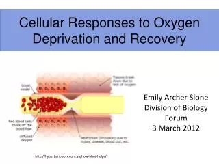
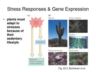
Cellular Responses To Stress. Normal Cell. Reversible injury. Adaptation. Cell Injury. Irreversible Injury. Necrosis. Apoptosis. ADAPTATION. Hypertrophy Hyperplasia Atrophy Metaplasia. Hypertrophy. Definition ? Which cell ? Example ?.

E N D
Normal Cell Reversible injury Adaptation Cell Injury Irreversible Injury Necrosis Apoptosis
ADAPTATION • Hypertrophy • Hyperplasia • Atrophy • Metaplasia
Hypertrophy • Definition ? • Which cell ? • Example ?
Physiologic hypertrophy of the uterus during pregnancy. A, Gross appearance of a normal uterus (right) and a gravid uterus (left)
B, Small spindle-shaped uterine smooth muscle cells from a normal uterus. Compare this with (C) large, plump hypertrophied smooth muscle cells from a gravid uterus.
Hyperplasia • Definition ? • Which cell ? • Example ? • Relation to CA
Here is one of the nodules of hyperplastic prostate, with many glands along with some intervening stroma. The cells making up the glands are normal in appearance, but there are just too many of them.
The prominent folds of endometrium in this uterus opened to reveal the endometrial cavity are an example of hyperplasia.
Atrophy • Definition ? • Causes with Examples ?
The testis at the right has undergone atrophy and is much smaller than the normal testis at the left.
This is cerebral atrophy in a patient with Alzheimer disease. The gyri are narrowed and the intervening sulci widened.
There are some muscle fibers here that show atrophy. The number of cells is the same as before the atrophy occurred, but the size of some fibers is reduced.
Metaplasia • Definition ? • Columnar to Squamous ? • Squamous to Columnar ? • Connective Tissue Metaplasia • Relation to CA
Metaplasia of normal columnar (left) to squamous epithelium (right) in a bronchus, shown (A) schematically and (B) histologically.
Metaplasia of laryngeal respiratory epithelium has occurred here in a smoker. The chronic irritation has led to an exchanging of one type of epithelium (the normal respiratory epithelium at the right) for another (the more resilient squamous epithelium at the left).
Metaplasia of the normal esophageal squamous mucosa has occurred here, with the appearance of gastric type columnar mucosa.
ApoptosisThe apoptotic cells are enlarged, pink from loss of cytoplasmic detail, and without nuclei
In this fetal thymus there is involution of thymic lymphocytes by the mechanism of apoptosis.
Types Of Necrosis • Coagulative Necrosis • Liquefactive Necrosis • Caseous Necrosis • Gangrenous Necrosis • Fibrinoid Necrosis • Fat Necrosis
Here is myocardium in which the cells are dying. The nuclei of the myocardial fibers are being lost. The cytoplasm is losing its structure, because no well-defined cross-striations are seen.
Two large infarctions (areas of Coagulative necrosis) are seen in this sectioned spleen.
This is the typical pattern with ischemia and infarction (loss of blood supply and resultant tissue anoxia)in the renal cortex of the kidney.
The contrast between normal adrenal cortex and the small pale infarct is good.
The liver shows a small abscess here filled with many neutrophils. This abscess is an example of localized Liquefactive necrosis.
This is Liquefactive necrosis in the brain in a patient who suffered a "stroke" with focal loss of blood supply to a portion of cerebrum. This type of infarction is marked by loss of neurons and neuroglial cells and the formation of a clear space at the center left.
Grossly, the cerebral infarction at the upper left here demonstrates Liquefactive necrosis. Eventually, the removal of the dead tissue leaves behind a cavity.
This is fat necrosis of the pancreas. Appear grossly as the soft, chalky white areas seen here on the cut surfaces.
This is the gross appearance of Caseous necrosis in a hilar lymph node infected with tuberculosis.
Microscopically, Caseous necrosis is characterized by acellular areas, as the tissue architecture is completely lost (at the upper right)
This is gangrene of the lower extremity. This patient had diabetes mellitus.
Here is fatty change of the liver due to accumulation of lipid in the cytoplasm of hepatocytes.
The brown coarsely granular material in macrophages in this alveolus is hemosiderin that has accumulated as a result of the breakdown of RBC's and release of the iron in heme. The macrophages clear up this debris, which is eventually recycled.
The sclera of the eye is yellow because the patient has jaundice
This is dystrophic calcification in the wall of the stomach. At the far left is an artery with calcification in its wall. There are also irregular bluish-purple deposits of calcium in the submucosa.
A 45 year old woman is investigated for hypertension and found to have enlargement of the left kidney. The right kidney is smaller than normal. Contrast studies reveal stenosis of the right renal artery.The Size Change In Both Kidney Is An Example Of Which Adaptive Change ?