Download
1 / 42
640 likes | 2.1k Vues
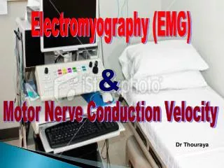
ELECTROMYOGRAPHY AND MOTOR NERVE CONDUCTION VELOCITY. ELECTROMYOGRAPHY (EMG). It’s a recording of electrical activity of the muscle by inserting needle electrode in the belly of the muscles or by applying the surface electrodes.

E N D
ELECTROMYOGRAPHY (EMG) • It’s a recording of electrical activity of the muscle by inserting needle electrode in the belly of the muscles or by applying the surface electrodes. • The potentials recorded on volitional effort are derived from motor units of the muscle, hence known as motor unit potentials (MUPs).
Electromyography (EMG) is a technique for evaluating and recording physiologic properties of muscles at rest and while contracting.
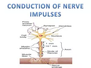
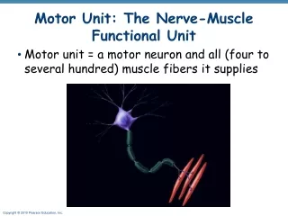
A motor unit is defined as one motor neuron and all of the muscle fibers it innervates.
Motor nerve conduction velocity of peripheral nerves may be closely correlated to their functional integrity or to their structural abnormalities. Based on the nature of conduction abnormalities two principal types of peripheral nerve lesions can be identified: Axonal degeneration and segmental demyelination. Motor nerve conduction velocity
In the patients of muscular weakness, muscle atrophy, traumatic or metabolic neuropathy, these tests are considered as an extension of the physical examination rather than a simple laboratory procedure.
OBJECTIVES At the end of the session the students should be able to: • Acquire a skill to perform the test by themselves. • Analyze the motor unit potentials and states their uses in health and diseases. • Determine and calculate motor conduction velocities of the peripheral nerves.
Requirements • Machine. • Electrodes. • Electrode jelly • Adhesive tape • Saline & antiseptic (70 % alcohol)
Instrument set up EMG • Sweep time 10msec / cm • Amplitude 1µV / cm • Audioamplifier on
Instrument set up Nerve conduction velocity • Sweep time 2msec / cm • Amplitude 1µV / cm • Stimulator set up • Frequency 1 / sec. • Duration 0.2 msec. • Intensity gradually increasing (MAM)
Procedure EMG • Select a volunteer and explain him the procedure. • Put the ground electrode over the forearm after soaking with saline. • Clean the skin over the selected muscle. • Apply the surface electrodes with the electrode jelly and reference electrode over bony point at least 3 cm apart.
Cont… • Put the sweep run (continuous). • Ask the subject to relax to evaluate any resting activity. • Ask the subject to exert mild voluntary effort then moderate effort while continue recording. • Change the sweep speed to 100msec/cm and then ask the subject to exert maximum effort to determine interference pattern.
Analysis EMG • Spontaneous activity • The skeletal muscle is silent at rest, hence spontaneous activity is absent.
Normal MUPs • Bi – Triphasic • Duration – 3 – 15 mSec. • Amplitude – 300μV – 5 mV
Abnormal MUPs In neurogenic lesion or in active myositis, the following spontaneous activity is noted • Positive sharp wave: • A small potential of 50 to 100 µV, 5 to 10 msec duration with abrupt onset and slow outset.
Positive SharpWaves Fibrillation Potentials
Fibrillation potential: • these are randomly occurring small amplitude potentials or may appear in runs. The audioamplifier gives sounds, as if somebody listen sounds of rains in a tin shade house. These potentials are generated from the single muscle fiber of a denervated muscle, possibly due to denervation hypersensitivity to acetyl choline.
Fasciculation potentials: • These are high voltage, polyphasic, long duration potentials appear spontaneously associated with visible contraction of the muscle. They originate from a large motor unit which is formed due to reinnervation of another motor unit from the neighboring motor unit.
EMG: Spontaneous Activity Fasciculation Potential
Typical MUAP characteristics in myopathic, neuropathic & normal muscle
Nerve Conduction studies • A nerve conduction study (NCS) is a test commonly used to evaluate the function, especially the ability of electrical conduction, of the motor and sensory nerves of the human body. Nerve conduction velocity (NCV) is a common measurement made during this test.
Procedure for MNCV • Give assurance to the subject about the short harmless electric stimulation. • Adjust the sweep speed to 2msec / cm. • Adjust stimulus duration to 0.2 msec and stimulus frequency to 1 / sec. • Apply electrode jelly on plate electrode.
Cont… • Put recording electrode over the thenar eminence for median nerve conduction velocity. • Fix the reference electrode 3 cm away & over a boney point.
Cont.. • Soak the stimulating electrode with saline and put it over median nerve at elbow. • Increase the stimulus intensity in steps. In each step give stimulation manually by pressing the stimulation switch once or twice until a visible muscle contraction is seen and a reproducible compound action potential (CMAP) is recorded. • Store the CMAP in the first channel.
Cont… • Change the stimulating site i.e. from elbow to wrist. • Stimulate the nerve & record the CMAP for median nerve at wrist. • Measure the distance from elbow to wrist with a measuring tape. • Measure the latency in first CMAP & in the next CAMP. • Enter the distance between the elbow and wrist.
MNCV • MNCV will appear. • It can also be calculated by formula • MNCV (m/sec)= • l1 = latency at elbow. • l2 = latency at wrist
Amplitude Duration Analysis of MNCV
Distance d = 284 mm Latency At elbow L1 = 8.5 ms Latency At wrist L2 = 3.5 ms
Normal values for conduction velocity • In arm • 50 – 70 m / sec. • In leg • 40 – 60 m / sec.