Download
1 / 48
630 likes | 2.86k Vues
ELECTROMYOGRAPHY AND MOTOR NERVE CONDUCTION VELOCITY. Dr. Salah Elmalik. Objectives. At the end of the lecture student should be able to: Define nerve conduction study (NCS) and electromyography ( EMG) . Explain the procedure of NCS

E N D
ELECTROMYOGRAPHY AND MOTOR NERVE CONDUCTION VELOCITY Dr. Salah Elmalik
Objectives At the end of the lecture student should be able to: • Define nerve conduction study (NCS) and electromyography ( EMG) . • Explain the procedure of NCS • Define the normal conduction velocity in upper limb and lower limb nerves . • Define the motor unit potentials ( MUPs) and how they are changed in muscle and nerve diseases .
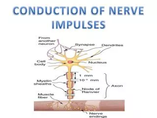
Nerve Conduction studies • A nerve conduction study (NCS) is a test commonly used to evaluate the function, especially the ability of electrical conduction, of the motor and sensory nerves of the human body. • Nerve conduction velocity (NCV) is a common measurement made during this test.
Motor nerve conduction velocity of peripheral nerves may be closely correlated to their functional integrity or to their structural abnormalities. Based on the nature of conduction abnormalities two principal types of peripheral nerve lesions can be identified: Axonal degeneration and segmental demyelination. Motor nerve conduction velocity
In the patients of muscular weakness, muscle atrophy, traumatic or metabolic neuropathy, these tests are considered as an extension of the physical examination rather than a simple laboratory procedure.
MOTOR CONDUCTION STUDIES • The The recorded potential, known as the compound muscle action potential (CMAP), represents the summation of all underlying individual muscle fiber action potentials. • CMAP is a biphasic potential with an initial upward deflection from the baseline • For each stimulation site : the latency, amplitude, duration, of the CMAP are measured . • A motor conduction velocity can be calculated after two sites of stimulation, one distal and one proximal.
MCS Procedure • The active recording electrode is placed on the center of the muscle belly (over the motor endplate), and the reference electrode is placed distally about 3-4 cm • The stimulator then is placed over the nerve that supplies the muscle.
Cont. • As current is slowly increased from a baseline: more of the underlying nerve fibers are brought to action potential, and subsequently more muscle fiber action potentials are generated. • most nerves require a current in the range from 20 to 50 mA to achieve supramaximal stimulation.
When the current is increased to the point that the CMAP no longer increases in size, one presumes that all nerve fibers have been excited and that supramaximal stimulation has been achieved. The current then is increased by another 20% to ensure supramaximal stimulation.
LATENCY • Latency measurements usually are made in milliseconds (ms). • The latency is the time from the stimulus to the initial deflection from baseline
Conduction Velocity • It’s measurement of the speed of the conducting nerve axons • It is calculated by dividing the change in distance (between proximal stimulation site & distal stimulation site in mm) by the change in time (proximal latency in msec minus distal latency in ms)
Normal values for conduction velocity • In arm • 50 – 70 m / sec. • In leg • 40 – 60 m / sec.
Amplitude • it is most commonly measured from baseline to the peak (baseline-to-peak) and less commonly from the first upward peak to the next downward peak (peak-to-peak).
CMAP amplitude reflects the number of muscle fibers that depolarize. • low CMAP amplitudes most often result from loss of axons (as in a typical axonal neuropathy) • average CMAP amplitude 3 mv
Duration • This is measured from the initial deflection from baseline to the final return • Duration characteristically increases in conditions that result in slowing of some motor fibers (e.g., in a demyelinating lesion).
MNCV • MNCV can also be calculated by formula • MNCV (m/sec)= • l1 = latency at elbow. • l2 = latency at wrist
Distance d = 284 mm Latency At elbow L1 = 8.5 ms Latency At wrist L2 = 3.5 ms
Patterns of nerve conductionNormal study of median nerve. • Note the normal median distal latency (DL) 3 ms, amplitude >4 m V, and conduction velocity (CV) >49 mls.
Axonal loss. • amplitudes decrease • CV is normal or slightly slowed. • DL is normal or slightly prolonged. • The morphology of the potential does not change between proximal and distal sites.
Demyelinationassociated with inherited disorders • CV is markedly slowed < 75% lower limit of normal) • DL is markedly prolonged (>130% upper limit of normal). • However, there usually is no change in configuration between proximal and distal stimulation
ELECTROMYOGRAPHY (EMG) • It’s a recording of electrical activity of the muscle by inserting needle electrode in the belly of the muscles or by applying the surface electrodes. • The potentials recorded on volitional effort are derived from motor units of the muscle, hence known as motor unit potentials (MUPs).
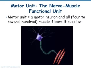
A motor unit is defined as one motor neuron and all of the muscle fibers it innervates.
Analysis EMG • Spontaneous activity • The skeletal muscle is silent at rest, hence spontaneous activity is absent.
Normal MUPs • Bi – Triphasic • Duration – 3 – 15 mSec. • Amplitude – 300μV – 5 mV
MUPs (2) During Mild Effort During Full Voluntary Effort . There is full recruitment ( you can not see the baseline ) During Moderate Effort note recruitment of additional motoneurons
Abnormal MUPs In neurogenic lesion or in active myositis, the following spontaneous activity is noted • Positive sharp wave: • A small potential of 50 to 100 µV, 5 to 10 msec duration with abrupt onset and slow outset.
Positive SharpWaves Fibrillation Potentials
Cont… • Fibrillation potential: • these are randomly occurring small amplitude potentials or may appear in runs. The audioamplifier gives sounds, as if somebody listen sounds of rains in a tin shade house. These potentials are generated from the single muscle fiber of a denervated muscle, possibly due to denervation hypersensitivity to acetyl choline.
Cont… • Fasciculation potentials: • These are high voltage, polyphasic, long duration potentials appear spontaneously associated with visible contraction of the muscle. They originate from a large motor unit which is formed due to reinnervation of another motor unit from the neighboring motor unit.
EMG: Spontaneous Activity Fasciculation Potential
Typical MUPs characteristics in myopathic, neuropathic & normal muscle