Download
1 / 25
250 likes | 279 Vues
Explore the characteristics, properties, and structures of cellulosic fibres like cotton and bast fibres. Understand the molecular compositions, crystallinity, and manufacturing processes in detail. Learn about wool and silk fibre structures for a comprehensive insight.

E N D
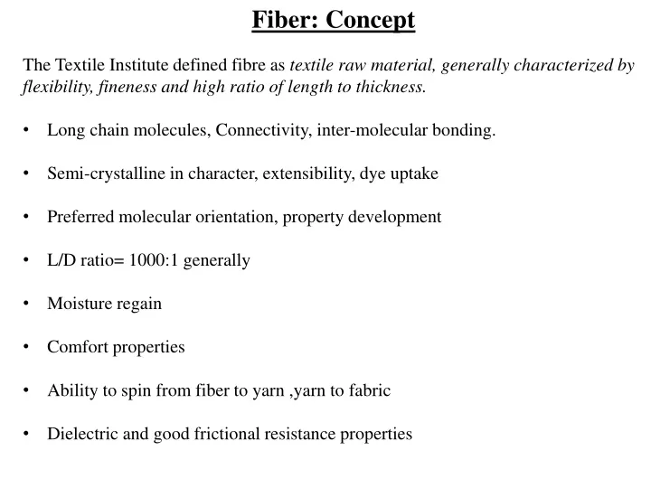
Fiber: Concept • The Textile Institute defined fibre as textile raw material, generally characterized by flexibility, fineness and high ratio of length to thickness. • Long chain molecules, Connectivity, inter-molecular bonding. • Semi-crystalline in character, extensibility, dye uptake • Preferred molecular orientation, property development • L/D ratio= 1000:1 generally • Moisture regain • Comfort properties • Ability to spin from fiber to yarn ,yarn to fabric • Dielectric and good frictional resistance properties
CELLULOSIC FIBRE • Cellulose is the most abundant polymer occurring in nature. dry wood consists of 40–55% cellulose, 15–35% lignin and 25–40% hemicelluloses. • It is the structural material in plants and also occurs in certain bacteria and some marine organisms. • In cotton fibres, it is found in an almost pure form. • Glucose, C6H12O6, has a ring structure of one oxygen –O– atom and five –CH– groups, four with pendant –OH groups and one with pendant –CH2OH . • The rings are not flat but bent in a chair configuration and each successive unit is rotated through 180° with respect to the molecular axis.
For the formation of cellulose within plant cells, glucose molecules come to enzyme complexes and add on successively with the elimination of water to form the long polymer molecules. • Irregular conformations with distributed hydrogen bonds are found in amorphous regions of regenerated fibres. • The molecule is unbranched and unfolded. (a) The polymer;(b) crosslinking by hydrogen bonds; (c) crystal;
It is estimated that the natural cellulosic chains contain about 104 glucose rings, giving a length of 5 μm and a width of 8 × 10⁻⁴ mm. • There are a number of active sites on the enzyme complexes so that about 30 cellulose chain molecules form together and can crystallize as fibrils. • The crystalline form, it is known as cellulose I and has all the chains running in the same directions. Two-thirds crystalline. • The fibrils are then laid down in helical layers to form the cell walls of plants. • In dry fibres, there is hydrogen bonding between the fibrils, but this reduces as water is absorbed with a consequent reduction in mechanical stiffness. • When cellulose is dissolved (or during swelling in caustic soda in mercerization), the parallel arrangement of molecules is lost. Recrystallization adopts a preferred cellulose II form with anti-parallel chains.
Cotton Fibres Diameter 12 to 20 μm , Single Cell • In the living seed cells, cellulose molecules grow by the addition of glucose molecules at enzyme complexes. • Virgin polymerization. • Thirty molecules form at each enzyme complex and assemble into fibrils, which are the building blocks of the structure. • First, a primary wall forms with the ultimate dimensions of the fibre. • The bulk of the material is laid down in helical layers as a secondary wall. • At maturity, there is still an open lumen at the centre of the fibre, which collapses to give a kidney-shaped fibre cross-section. Helical – ribbon shape, reversal spiral ~ same for all layers, helix angle 30o, 67% crystallinity
Cotton Fibres Chain Details: D.P = +10,000; Molecule is ribbon shapedRigidity and flexibility, Extensive Hydrogen Bonding Bast Fibres Flax, Hemp, Jute.i) Multicellularii) Less celluloseiii) Helix angle 7- 15o Cellulose Acetate -OH group replaced by CH3COO-Secondary Acetate – 1 to 2 groups are replaced. Less crystalline due to bulky side group and –OH groups. Weak and Extensible.Triacetate – 3 groups are replaced by acetate. 40% crystalline,greater dimensional stability, crease resistance.
Wool Fibre Structure • The fibres grow from follicles in the skin. • They are composed of many elongated cells linked by a cell membrane complex, which contains peptides. • Keratin protein is laid down in sequence as the cell grows. • Keratin crystallizes in an α-helical lattice, which under tension opens to extended β-sheets. • The molecules assemble in a sequence of twisted pairs to form fibrils containing 32 molecules. • Proteins form in the outer cells to give a multi-layer cuticle. • The core of the fibre is divided into an ortho- and para-cortex, with differences in the composition of the keratin associated proteins (KAPs). • The two halves contract differently on drying, causing wool to develop crimp. • The cuticle has a structure of scales, which are like an escarpment with steep faces towards the tip and gentle slopes to the root. • 25-30% crystallinity, Fringed fibril structure
Silk Structure • Simpler protein block copolymers, which form within gland cells. • The process is similar to solution spinning of manufactured fibres, but with important difference. • Virgin polymerization. Heighest orientation Sheath core structure. Outer layer coagulates first, lower time for orientation, followed by middle and nextthe inner layer. Mediumorientation Low orientation Fibre cross section Fibrillar Structure 65-70% crystalline.
Slow growth, cm/minute for silks or cm/month for cotton and wool compared with km/minute in industrial production, and genetic control lead to complex specificity in fibre structures that cannot be matched in commercial manufacturing. Requirements of fibreforming polymers • Long-chain molecules • Amore or less parallel arrangement of the molecules • Lateral forces to hold the molecules together and give cohesion to the structure. • Freedom of molecular movement. • Openness
Historical view of fibre fine structure • Nageli suggested in the middle of the nineteenth century that cell membranes of plant fibres are composed of sub-microscopic crystallites, called micelles, embedded in a matrix of some indeterminate inter-micellar substance. • In 1920s it was accepted that atoms or groups of atoms could add on indefinitely to form macromolecules, polymers composed of many monomers, with a range of molecular weights. • By then X-ray diffraction had confirmed that the fibres contained both crystalline and amorphous material. • Seifriz (1929) and Meyer (1930) modified Nageli’s hypothesis by showing the micelles as composed of polymers, but still separated by an indeterminate matrix,
Molecular weight determinations then showed that the polymer molecules are about ten times longer than the length of crystallites estimated from the X-ray diffraction. • This led Gerngross and Herrmann to propose a fringed micelle structure, in which polymer molecules passed in and out of crystallites through an amorphous matrix. • One feature that is common to all the drawings from this period is that the molecules run continuously along the oriented fibre structures with no folding back.
Staudinger (1932) suggested that the structure was a continuous imperfect crystal in which the ends of the molecules acted as local defects, • Howsmon and Sisson (1954) pointed out that there could be a whole spectrum of increasing order from a fully random arrangement to perfect crystals.
For manufactured fibres, the laboratory studies of polymer crystallization led to a change of view of fibre structure, particularly for melt-spun nylon and polyester fibres. • Slow crystallization of polymer melts or solutions starts from a small nuclei. • crystallization proceeds there by repeated branching until crystal growth is equal in all directions. • Spherulites form. • At the centre of spherulites, the nuclei are present as sheaf-like assemblies of parallel molecular segments fringing off into the first branches and fringed micelle structure forms.
More surprising was Keller’s discovery of single crystals of polyethylene in which the molecules folded back and forth at the ends of the crystal. This led to a modified fringed micelle model. • At the ends of the crystallites, there was a mixture of chain folding and fringing into tie-molecules, which led to other crystallites.
In natural fibres with stiffer molecules, electron microscopy showed that the structure was fibrillar and not micellar. • In cotton, the structure was in one sense wholly crystalline: crystalline fibrils laid down in a helical array in the cell walls. • Cotton is two-thirds crystalline and one-third amorphous resulted from a lack of register between fibrils with a diameter of about 3 nm. • In wool, keratin fibrils of 7 nm diameter were spaced 10 nm apart in a matrix of KAPs. • Hearle proposed a fringed-fibril structure for natural fibres. • This combines (a) a fibrillar form and (b) the ideas inherent in the fringed-micelle structure of distinct crystalline and non-crystalline regions with chain molecules running continuously through each type of region. • Polymers such as polyethylene have been found to crystallise in lamellar forms linked by tie-molecules.
Forming of Spherullite structure at bulk polymerization: Acrylic bulk polymerization
Alternative, more uniform structures, such as paracrystalline, crystals with distributed defects, or amorphous with correlation, were also proposed.
Parameters for quantitative description of fibre structure Degree of order, which is frequently referred to as crystallinity. Degree of localization of order, which differentiates between structures with a well-defined separation into crystalline and amorphous regions and more uniform structures described as paracrystalline, as amorphous with correlation, or as containing distributed crystal defects. Length/width ratio of localized units, which distinguishes between lamellar, micellar and fibrillarforms. Degree of orientation, which may be separated into crystalline and amorphous orientation and may include transverse as well as axial orientation. Size of localized units, which may include cross-sectional shape. Molecular extent, which is an inverse measure of the degree to which molecules are folded to-and-fro, either at the ends of crystallites or in amorphous regions, or alternatively are more or less fully extended along the fibre.
Degree of order would be theoretically defined as the mean value of some correlation function relating the position of neighboring chains. Practically, it could be defined in terms of density by the expression: Where, ρ is the fibre density, ρam the density of amorphous (non-crystalline) material and ρcr the density of crystalline material Degree of localization of order would be theoretically defined by some measure of the spread of values of degree of order taken over zones a few molecules wide. The length/width ratio of the units is a more straightforward parameter, ranging from infinity for very long fibrils down to unity for cubic micelles.
Degree of orientation, defined theoretically by a mean angle between the chain molecules and the fibre axis. Size of localized units indicate the difference between a coarse and a fine texture. Molecular extent, is an inverse measure of the degree to which molecules are folded to-and-fro, either at the ends of crystallites or in amorphous regions, or alternatively are more or less fully extended along the fibre.
Order, orientation and extent A perfectly ordered structure must be perfectly oriented with fully extended chains, and a completely disordered structure must be completely disoriented with short fold lengths.