Download
1 / 31
310 likes | 528 Vues
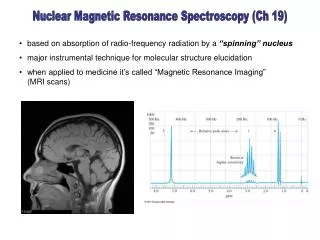
Nuclear Magnetic Resonance Spectroscopy (Ch 19). based on absorption of radio-frequency radiation by a “spinning” nucleus major instrumental technique for molecular structure elucidation when applied to medicine it’s called “Magnetic Resonance Imaging” (MRI scans). Theory of NMR.

E N D
Nuclear Magnetic Resonance Spectroscopy (Ch 19) • based on absorption of radio-frequency radiation by a “spinning” nucleus • major instrumental technique for molecular structure elucidation • when applied to medicine it’s called “Magnetic Resonance Imaging” (MRI scans)
Theory of NMR Nuclear Spin States • certain nuclei possess properties that can be explained by assuming that they are spinning (precessing) about an axis (like a top) • these nuclei possess a spin quantum number (I), e.g I = 3/2 for 11B (see table) • the nucleus possesses energy levels - spin states – that are quantized. • The maximum number of spin states is equal to 2I + 1, and each spin state has a magnetic quantum number mI that ranges from - mI to + mIin steps of one, e.g.11B • these different spin states are “equal-energy” in the absence of an external magnetic field, but split in a magnetic field • Only those nuclei with an odd mass number, an odd atomic number, or both - may possess spin http://hyperphysics.phy-astr.gsu.edu/hbase/mechanics/imgmech/topp.gif
mI = -1/2 mI = +1/2 high potential energy: aligned against field mI = -1/2 1H low potential energy: aligned with field mI = +1/2 Bo Energy Levels in a Magnetic Field The nucleus is a charged "particle" and its spin will induce a magnetic moment () - If apply an external magnetic field Bo, then the spin states will split and the magnetic moments will align themselves either with or against the applied magnetic field -
e.g. nuclear spin states for 11B - mI = -3/2 mI = -1/2 11B mI = +1/2 Bo mI = +3/2
after absorption of the photon, the nucleus “flips” its spin to the higher energy spin state Splitting of Spin States & Absorption of Radiation The magnitude of the energy level splitting increases with the strength of the external magnetic field, e.g. for 1H - mI = -1/2 E E = hphoton mI = +1/2 Bo
from the Quantum Mechanics of 1H, a given spin state energy is given by - • = “magnetogyric ratio” mI = magnetic quantum number h = Planck’s constant Bo = external magnetic field strength The energy gap between spin states is equal to - And the frequency of the absorbed photon is -
Example 19-1 Many proton NMR instruments employ a magnet that provides a field strength of 4.69 Tesla. At what frequency would the hydrogen nucleus absorb in such a field?
Classical Description of NMR Precession of Nuclei in a Field The nucleus precesses at the Larmor Frequency which must be the same as the frequency of the absorbed photon -
Circularly Polarized Radiation Before the radio-frequency photon can be absorbed, it must be circularly polarized. The rotational rate of the circularly-polarized beam must match the nucleus precessional frequency (Larmor Frequency).
Fourier Transform NMR The sample is hit by pulses of radio-frequency radiation (100 – 1000 MHz)
Pulsed Excitation B1 When the nucleus relaxes to the ground state, it emits the FREE INDUCTION DECAY (FID) signal
Free Induction Decay (FID) The FID consists of the superposition of all frequencies emitted by the protons in the molecule. Protons that are in different chemical environments emit slightly different frequencies from the Larmor Fequency. The NMR spectrum is obtained from the FID via Fourier Transformation
Origin of the Chemical Shift Protons on different parts of a molecule experience different chemical environments because of “shielding” and “deshielding” - circulating electrons in the molecule induce an internal field that can align either with or against the applied magnetic field. Shielding and deshielding can be influenced by electronegativity effects (withdrawal of electron density).
F Cl H H H H H H • not all protons in the 4.6 Tesla magnetic field will precess at 200 MHz because of shielding effects • the net magnetic field that the nucleus experiences will be increased or decreased by the internal induced manetic field protons slightly more deshielded Bo precesses at 1 precesses at 2 1 - 2 200 Hz out of the applied field of 200 MHz
The chemical shift is measured relative to a standard – TetraMethylSilane (TMS) – protons are more shielded than in most molecules deshielded shielded
Br F H I Cl H H H H H H H H H H H H H H H Theory of the Chemical Shift A. Electronegativity Effects increases as the electronegativity of an attached atom increases. = 4.26 = 3.05 = 2.68 = 2.16 = 0.23
B. Hybridization Effects 1s 2s 3s 4s 2p Probability of finding the electron → 3p 4p 3d 4f 4d distance from nucleus → http://www.pha.jhu.edu/~rt19/hydro/img73.gif
D. Magnetic Anisotropy Aromatic Protons
deshielded shielded Bo "1st Order" Interpretation of NMR Spectra Spin-Spin Splitting or Coupling (N+1 Rule) Number of peaks = number of nearest neighbors + 1
FT-NMR Spectrometers • Pulser Switch • Magnets • Frequency Locking • Shimming • Sample Spinning
transmitter-receiver coils RF Transmitter RF Amplifier N S shimming coils shimming coils
Resolution of FT-NMR Spectrometers As in FTIR where resolution depended on sampling the interferogram at the Nyquist frequency, in FT-NMR the FID signal must be sampled at a rate proportional to the resolution.
npts = 2 x range resol. 400 Hz “aliased” frequency 1600 Hz signal Nyquist Theorem:A periodic signal must be sampled twice per period (I.e. sampled at twice the frequency of the signal e.g. A resolution of 1 Hz is desired using a 25 MHz NMR spectrometer. The scan range is 200 ppm. How many points need to be sampled? resol in ppm, = 1 Hz/25 MHz = 0.040 ppm npts = 2(200 ppm)/(0.040 ppm) = 10,000 pts (example from “Chemical Instrumentation”, 2nd ed, by Howard A. Strobel, 1973, Addison-Wesley) FID Signal
Carbon-13 NMR • Structural information about the carbon skeleton of the molecule • low isotopic abundance of 13C = 1.1% • low probability of having neighboring 13C atoms, so no spin-spin coupling = simplified spectrum • low isotopic abundance results in weak signals, but S/N improved by signal averaging (the Multiplex Advantage again) • 13C and 1H have the same spin quantum number (1/2), so there is spin-spin coupling between carbons and attached protons. Eliminated by using a spin decoupler.


![G3 - RADIO WAVE PROPAGATION [3 Exam Questions -- 3 Groups]](https://cdn0.slideserve.com/819386/g3-radio-wave-propagation-3-exam-questions-3-groups-dt.jpg)