Download
1 / 38
390 likes | 700 Vues
Misidentification and Contamination of cell lines 2009. BioSafety Office HSC 3N1C, ext. 24956 Complete the quiz and return to HSC 3N1C or fax 905 528 8539 withyman@mcmaster.ca cassidk@mcmaster.ca.

E N D
Misidentification and Contamination of cell lines 2009 BioSafety Office HSC 3N1C, ext. 24956 Complete the quiz and return to HSC 3N1C or fax 905 528 8539 withyman@mcmaster.cacassidk@mcmaster.ca
introductory information (2-4)quality of cell lines (6-12) cell line identification (13-18) contamination (19- 37)summary (38)
Objective of this presentation To increase awareness of the need for verification of the identity of the cell lines / organisms being used for research The Basics 1) establish a strict code of practice for your lab 2) ensure everyone adheres to it
Testing for contamination or misidentification of cell lines “Evidence suggests that up to one-third of tumor cell lines being used in scientific research are affected by inter- or intraspecies cross contamination or have been wrongly identified, thereby rendering many of the conclusions doubtful if not completely invalid.” Lancet Oncology, vol. 2, July 2001, p. 393 The following is, in part, from ATCC informationposted on line
General Accessioning Processfor commercial cell resource centers >>>>>> Token freeze • Contamination checks >>>>>>>Verification by depositor • Species verification • Post-freeze recovery • Post-freeze viability • Growth curve • Characterization tests >>>>> Seed stock>>>>>> Distribution • Contamination checks Contamination checks • Species verification Species verification • Post-freeze recovery Post-freeze recovery • Post-freeze viability Post-freeze viability • Characterization tests Characterization tests
Experimental success corresponds directly to the quality and conditions of cell lines used in practice…what tests do you use to confirm cell line identification?
To maintain high cell culture standards and ensure reliable, reproducible results, the use of authenticated and quality-tested cell lines is highly recommended In practice: where can you find the protocol for routine cell culture standards in your lab?
Cells that are kept too long in culture and are not periodically tested for genotypic or phenotypic stability may no longer be reliable models of the original source material. In practice: do you keep records of cell passages? In practice: what are the indicators for your cell lines?
Cellular morphology can vary between lines depending on the health of the cells and, in some cases, the differentiation state. Morphology can change with plating density as well as with different media and sera. Cells may develop permanent changes if grown in sub-optimum conditions such as temperature, gas concentration or pH variability.
Avoid the use of cell lines that have been in culture too long as a first step to ensuring reliable and reproducible results. Important characteristics can permanently change when cells are cultured for extended periods or are exposed to culture media extreme conditions.
High-passage cell lines can exhibit alterations in the following properties: • morphology • growth rates • response to stimuli • protein expression and signaling
For more information on high passage cell lines read Bulletin #6 and #7 on this web page.http://fhs.mcmaster.ca/safetyoffice/biosafety_cell_line.html
Assessing cell identityis recommended: 1) when a cell line is first isolated 2) at regular intervals ie new bulk batch 3) when irregular results are experienced
PCR Use DNA primers to test for unique cellular sequences In practice: one vial should be tested when freezing down or recovering a cell line In practice: available in many labs To be done: good identity charts are being developed
Surface markers/flow cytometry Use of antibodies with a fluorescent tag specific to cellular proteins to identify cell type • In practice: for selected valuable cells due to the expense, however, the results may not be unique to one cell type
Karyotyping 1) is performed to identify the species 2) karyotyping is a basic and indispensable test to determine if the line has maintained a stable genotype In practice: infrequently done in a routine research lab
Short tandem repeat profiling 1) establishes a DNA fingerprint for human cell lines. 2)STR profiling uses multiplex PCR to simultaneously amplify the amelogenin gene and eight of the most informative polymorphic markers in the human genome. 3) the pattern of repeats results in a unique STR identity profile for each cell line analyzed. 4) STR analysis is critical for verifying the identity of human cell lines.
Isoenzyme analysis 1) is used to verify the species of origin. 2) isoenzymes are differentiated based on electrophoretic properties. 3) can be used to verify information regarding the source species of a cell line and check for species cross contamination.
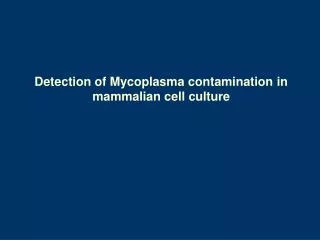
Contamination is a problem in cultures The problem and detection of contamination caused by aggressive cell lines, bacteria, mycoplasma, yeast, virus or fungus
Contamination by mycoplasma or other bacterial, viral or fungal agentscan seriously affect: • cell growth and function • transfection • morphology and differentiation state • gene expression
the damaging effects of mycoplasma contamination on cell lines is a major problem in cell culture. • the problem is exacerbated by the exchange of cell lines between labs. • because mycoplasma growth in cell cultures cannot be detected visually or under the microscope, routine testing remains the only assurance against contamination.
Mycoplasma contamination 1) may show as abnormal cell growth eg agglutination, decreased growth rate, reduced saturation. 2) cells may show abnormal labeling with 3H thymidine. 3) invisible under regular microscopy. 4) requires kits for testing.
5)Failure to raise specific cell surface protein antibodies would warrant mycoplasma testing of the culture. 6) test all continuous cell lines regularly ……….and document results. 7) if cell lines are contaminated, your research results are likely to be affected. 8) Some media may inhibit the growth of mycoplasma eg G418.
Tests to detect Mycoplasma 1) ELISA 2) 16 S rRNA - see Dr Andrews web site 3) Commercial kits –results may vary eg Stratagene Mycoplasms Plus PCR – has been found to have false negatives eg Lonza MycoAlert Assay control 4) DAPI
5) Fluorescent staining of DNA using Hoechst 33258 requires a fluorescent microscope Staining of the nuclei of cells are the positive control for this test May also detect bacterial contamination
6) Other in-house tests could be available depending on the type of work being done in a specific lab. - immunostaining - PCR - autoradiograph with 3H thymidine - southern blot probes for mycoplasmal DNA
Clearing Mycoplasmafrom cultures These are available in conjunction with commercial kits but there is no specific recommendation. Look for information on web sites for products such as: BM Cyclin Myco Zap Plasmacyte
Sourcesof viral contamination • Cell culture reagents of animal origin eg serum supplement • Technician/student operator • Tissues of animal origin used as isolation source • Cross contamination from another line
Potential effects of viral contamination • Lysis of host cell line • Persistent sub lethal infection • In a small number of cases, release of human infectious viral particles eg B95-8 or MT4 cells • Release of retrovirus-like particles eg CHO, BHK, murine myelomas
Avoiding contamination by viruses 1) buy only certified product from a known supplier 2) if possible, use primary culture sources from animals known to be free of viral contamination 3) use PCR for suspected viral contamination screening
Clearing of viral contamination discard any contaminated cultures, they cannot be cleaned.
Bacterial contamination Many cultures routinely use antibiotics (although not recommended), therefore, to test for bacterial contamination, it should be done following a period of antibiotic free culture to ensure that contaminants suppressed by antibiotics are not missed.
Quick check for differentiation of bacterial/yeast contamination Yeast tends to show as a yellow in phenol red media Bacteria tend to show as a pink in phenol red media
Checking for bacterial contamination Bacteria can often be seen under the light contrast microscope or with the use of staining techniques Cultures can be plated out for antibiotic sensitivity
Use of antibiotics for cell culture Antibiotics can be effective against bacteria, but may have toxic effects on cells. Antifungal agents may be more toxic to cell lines than antibiotics. Use of antibiotics should not become routine in a culture lab. Antibiotics can never be relied on as a substitute for good aseptic technique.
Cell line cross contamination When this occurs, it is often not recognized. The contaminating cell line is often very aggressive in its growth characteristics eg HeLa and overwhelms the existing line, therefore contamination is only suspected after unusual research results. Test for surface markers or species.
Cleaning a contaminated culture 1) best to discard the culture and all associated reagents and start fresh 2) attempt eradication of vital cultures only, with antibiotics, under experienced supervision, with post eradication quality controls 3) yeasts tend to be very resistant
In summary Keep good culture standards and records Test for cell characteristics regularly Be highly aware of changes At all times, support best practices in the use of cells, tissues and organisms in vitro