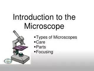
Introduction to the Microscope Lab Activity
370 likes | 407 Vues
Explore the world of microscopy in this lab activity where you will learn how to properly use and handle a compound light microscope, as well as make your own slides for observation. Discover the structure of plant cells through hands-on experiments.

Introduction to the Microscope Lab Activity
E N D
Presentation Transcript
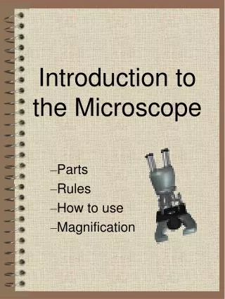
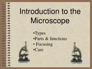
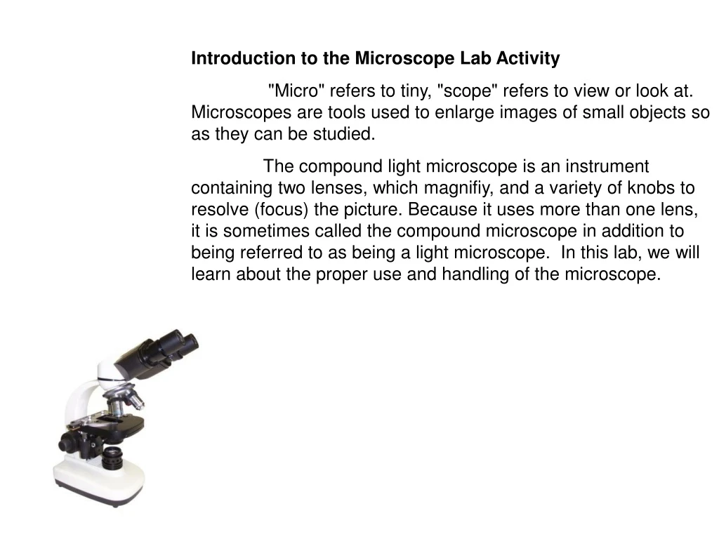
Introduction to the Microscope Lab Activity "Micro" refers to tiny, "scope" refers to view or look at. Microscopes are tools used to enlarge images of small objects so as they can be studied. The compound light microscope is an instrument containing two lenses, which magnifiy, and a variety of knobs to resolve (focus) the picture. Because it uses more than one lens, it is sometimes called the compound microscope in addition to being referred to as being a light microscope. In this lab, we will learn about the proper use and handling of the microscope.
Basic Rules for Caring for Microscopes • EVERYTHING on a microscope is unbelievably expensive, so be careful. • Hold a microscope firmly by the stand, only. Never grab it by the eyepiece holder, for example. • Hold the plug (not the cable) when unplugging the illuminator. • Since bulbs are expensive, and have a limited life, turn the illuminator off when not in use. • If used constantly on full power the bulb will overheat and blow (or gently melt the inside of the housing). This is not a good idea! • Always make sure the stage and lenses are clean before putting the microscope away. • NEVER use anything but good quality lens tissue on any optical surface, with appropriate lens cleaner or distilled water; organic solvents may separate or damage the lens elements or coatings. • Cover the instrument with a dust jacket when not in use. • Focus smoothly; don't try to speed through the focusing process or force anything. • If it isn't working DON'T try to fix it unless you really know what you are doing..
In botany – two kind of slides: • simpleslide – wet mount slide • prepared slide - can be stored for many years • Simple slide – materials needed: • Glass slide • Cover slip • Eye dropper • Beaker of water • Pincetes • Slices of gause • Blotting paper
How to make the microscope slide: • Carefully cut a very thin slice of the specimen using a scalpel - the thinner the slice, the easier it will be to view with your microscope. • Put one drop of water in the center of a plain glass slide - the water droplet should be larger than the slice of specimen. • Gently set the slice of specimen on top of the drop of water (pincetes might be helpful for this). If you are not able to cut a thin enough slice of the whole diameter of the specimen, a smaller section will work. • Take one cover slip and hold it at an angle to the slide so that one edge of it touches the water droplet on the surface of the slide. • Then, being careful not to move the specimen around, lower the cover slip without trapping any air bubbles beneath it. The water should form a seal around the sample. • Use the corner of a paper towel to blot up any excess water at the edges of the coverslip. • Begin with the lowest-power objective to view your slide. Then switch to a higher power objective to see more detail.
Introduction to the cell – cell forms Two types – parenchyma – polygonal, cubic or oval shape form – least specialised - prosenchyma - elongated or spindly shape form
Observation 1 - parenchyma cells of apple fruit • Methodology: • Add a few drops of water on the microscope slide • Crush a small piece of parenchyma tissuebetween glass slide and cover slip. Make sure there are no air bubbles. You can avoid them by lightly tappingof the tweezers.
Observation 2 – Prosenchyma cells in lower epiderma of onion leaf • Methodology: • Add a few drops of water on the microscope slide • Take a small piece of onion and peel off the membrane from the underside (the rough side). • Lay the membrane flat on the surface of the slide • Using a pin, lower the thin glass cover slip or cover glass onto the slide. Make sure there are no air bubbles.
Observation 3 – Cell structure - In rule : • Important structures inside the typical plant cell include: • Cell membrane - controls what substances enter and leave the cell • Cell wall - gives shape and support to the plant cell • Cytosol - medium in which all metabolic reactions occur • Nucleus - controls all activities of the cell • Nucleolus - makes RNA • Mitochondrion - carries out the reactions of respiration • Chloroplast - carries out the reactions of photosynthesis • Large central vacuole - stores food, water, minerals, vitamins, and wastes • Lysosome - destroys old, worn out cell organelles • Ribosome - makes protein The Microscope
Observation 3 – Cell organells in lower epiderma of onion leaf Materials needed: onion epiderma cells, jodine solution, ablatting paper - to blot up any excess water at the edges of the coverslip. The nuclous, cytoplasm and vacuole turn brown - there is no other organelles because the function forthat cells is a storage of carbohydrates and water that can be broken down and resorbed to supply the metabolic and growth needs of the plant.
Typical plant cell organelles: • Cell wall • Plastids • Vacuole
1.Cell wall - a tough, rigid and semi-permeable extracellular structure responsible for cell protection and support, is found in plants, bacteria, archaea, fungi and some protists. • Cell wall main functions may include: • - Structural and mechanical support • - Providing a chemically buffered environment • - Maintenance of cell shape • - Resistance to pressure of cell • - Rate and direction of growth control • - Regulate diffusion, a porous medium for circulation of water, minerals, and other small nutrients • - Rigid building blocks from which stable structures can be produced • - Storage of carbohydrates • - Protection against pathogens and other environmental factors • - Source of active signaling molecules • Cell to cell interactions
Structure components of cell wall – primary cell wall: It is located outside the cell membrane whose main function is to provide rigidity, strength, protection against mechanical stress and infection. Cell wall is made up of cellulose, pectins,glycoproteins, hemicellulose and lignin. The primary cell wall is characterized by a high proportion of pectin substances - 50-60%, cellulose - 30%, structural proteins - up to 10%. Rarely, it can participate, and lignin. It is characteristic of young cells in which the celulose is relatively small amounts. flexible and extensible and allowing the cell to grow.
However, the primary cell wall, can be defined as composed of cellulose microfibrils aligned at all angles. Microfibrils are held together by hydrogen bonds to provide a high tensile strength.
Gradually, the amount of cellulose and lignin increases at the expense of the pectin so that the mature cells in the ratio between the structural components is 35 - 50% cellulose, 25-30% lignin, 10-25 % hemicellulose, pectin - 0%. The outer part of the primary cell wall of the plant epidermis is usually impregnated with cutin and wax, forming a permeability barrier known as the plant cutucule
Incorporation the lignin between the fibers greatly reduces the mobility of fibers, the cell wall becomes thicker and stops growing - its formed secondary cell wall - a thick layer formed inside the primary cell wall after the cell is fully grown . The evolution of conducting tissues - xylem fibers, tracheids, and sclereids with rigid secondary cell walls was a critical adaptive event in the history of land plants, as it facilitated the transport of water and nutrients and allowed extensive upright growth. Secondary walls also have a major impact on human life, as they are a major component of wood and are a source of nutrition for livestock.
Features of cell division due to cellulose cell wall: In contrast to animal cell division in plants starting from the middle of the cell – from the midlle lamella. The middle lamella is laid down first, formed from the cell plate during cytokinesis, and the primary cell wall is deposited inside the middle lamella after that.
II. Typical cell organelles – Plastids Plastids is a major double-membrane organelle found, in the plants cells and algae. Plastids are the site of manufacture and storage of important chemical compounds – fatty acids, terpenes,and a starch, used by the cell. They often contain pigments used in photosynthesis, and the types of pigments present can change or determine the cell's color. For example, the components of the plant cuticule and its epicutucular wax are synthesized by the epidermal cells from palmitic acid , which is synthesized in the chloroplasts of the mesophyll tissues. All plastids are derived from proplastids, which are present in the meristematic regions of the plant. Proplastids and young chloroplasts commonly divide by binary fission, but more mature chloroplasts also have this capacity.
1.Chloroplasts • Chloroplasts‘- a green plastids, spread in all green part of the plants – leaves, buds, stems • The main role - to conduct photosynthesis with photosynthetic pigment chlorophyll, which captures the sunlight energy and converts it in the energy-storagemolecules ATP and NADPH. These molecules will used to organic molecules formation. Chloroplasts carry out a number of other functions - fatty acid synthesis, much amino acid synthesis, and the plants immune responce. The number of chloroplasts per cell varies from 1 in algae up to 100 in plants like Arabidopsis and wheat.
A cross section of a leaf, showing chloroplasts in its mesophyll cells. Stomal guard cells also have chloroplasts, though much fewer than mesophyll cells.
Chloroplasts - structure Like mitochondria, chloroplasts are surrounded by two membranes. The outer membrane is permeable to small organic molecules, whereas the inner membrane is less permeable and studded with transport proteins. The innermost matrix of chloroplasts, called the stroma, contains metabolic enzymes and multiple copies of the chloroplast genome. Chloroplasts also have a third internal membrane called the thylakoid membrane, which is extensively folded and appears as stacks of flattened disks in electron micrographs. The thylakoids contain the light-harvesting complex, including pigments such as chlorophyll, as well as the electron transport chains used in photosynthesis.
Observation 4 – Chloroplast in lower epidermis of Tradescantia virginica Materials needed: a leaf of Tradescantia virginica, Glass slide , cover slip, eye dropper , beaker of water
2. Chromoplasts - Chromoplasts are found in fruits, flowers, roots, and stressed and aging leaves, and are responsible for their distinctive colors. This is always associated with a massive increase in the accumulation of carotenoid pigments. The conversion of chloroplasts to chromoplasts in ripening is a classic example.
Function - Chromoplasts synthesize and store pigments such as orange carotene, yellow xanthophyllus, and various other red pigments. As such, their color varies depending on what pigment they contain. The main evolutionary purpose of chromoplasts is probably to attract pollinators or eaters of colored fruits, which help disperse seeds . However, they are also found in roots such as carrots and sweet potatous. They allow the accumulation of large quantities of water-insoluble compounds in otherwise watery parts of plants.
The pigments are cartenoids, a form of which is converted to vitamin A in the human digestive tract.(why you are suppose to eat carrots to improve your night vision: new findings indicate they can also help in the prevention of cataracts
Schematic representation of the main structural changes occurring during the chloroplast to chromoplast transition.
3. Leucoplasts - non-pigmented plastids in non-photosynthetic organs - roots,bulbs and seeds. • Functions - specialized for bulk storage of starch, lipid and proteins • - including the synthesis of fatty acids such as palmitic acid, many amino acids, and tetrapyrrole compounds. • Types • amyloplasts, • elaioplasts • proteinoplasts (also called aleuroplasts)
Amyloplasts are non-pigmented organelles found in some plant cells. They are responsible for the synthesis and storage of starch granules, through the polymerizationof glucose. Amyloplasts also convert this starch back into sugar when the plant needs energy. Large numbers of amyloplasts can be found in fruit and in underground storage tissies of some plants, such as in potato tubers.
Observation 6 – of starch granules (amyloplasts) of potato tubers and a banana fruit.
Elaioplasts are a type ofleucoplasts that is specialized for the storage of lipids in plants.Elaioplasts house oil body deposits as rounded plastoglobuli, which are essentially fat droplets. Elaioplasts in Persea americana (Lauraceae)
Proteinoplasts - for proteine storage and form the aleurone layer of maturing seed and tubers. Aleurone proteins can have two different morphological features - homogenous and heterogeneous. • the homogenous aleurone consists of similar protein bodies (e.g. Phaseolus vulgaris) • the heterogeneous aleurone consists of granules of different shapes and types of proteins covered with a membrane (e.g. Ricinus communis).
III. Vacuoles – a multifunctional organelles that are central to cellular strategies of plant development. • - share some of their basic properties with the vacuoles of algae and the lysosomes of animal cells. • lytic compartments, function as reservoirs for ions and metabolites, including pigments • involved in cellular responses to environmental and biotic factors that provoke stress. • In the plant vegetative organs act in combination with the cell wall to generate turgor, the driving force for hydraulic stiffness and growth. • In seeds and specialized storage tissues, they serve as sites for storing reserve proteins and soluble carbohydrates. Vacuoles - the essential parts of plant life
Vacuole – Most mature plant cells have one large vacuole that typically occupies more than 30% of the cell's volume, and that can occupy as much as 80% of the volume for certain cell types and conditions Plant cells use vacuoles to their advantage to sustain rapid cell enlargement during growth. Young cells typically possess many small vacuoles and ample cytoplasm. As cells mature, the small vacuoles merge to form a single, large vacuole, the contents of which are predominately water.
Plant cell vacuoles are bounded by a membrane – thetonoplast. Assume this membrane is similar to the plasma membrane with integral transport mechanisms. Scanning Electron Micrograph (SEM) of cut edge of a leaf, looking into cells which have been opened. Contents of the cells are seen through the thin membrane (the tonoplast membrane) which lines the large central vacuole of many plant cells.
Pigments - Anthocyanins and flavonoids located in the cell vacuoles are responsible for other colors of pigment. The anthocyanin-storing vacuoles of Rhoeo spathacea, a spiderwort, in cells that have plasmolyzed