Enterobacteriaceae Family: General Characteristics & Identification Methods
350 likes | 378 Vues
Learn about Enterobacteriaceae bacteria, normal flora in the intestine causing various diseases. Understand their unique characteristics, classifications, and identification methods such as MacConkey agar, EMB agar, and TSI agar. Discover E. coli infections and differentiation tests for this diverse bacterial family.

Enterobacteriaceae Family: General Characteristics & Identification Methods
E N D
Presentation Transcript
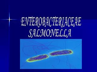
Lab 15 Enterobacteriaceae
Gram’s Negative Bacilli Enteric bacilli Non enteric bacilli Late-lactose fermenting (LLF): NLF after 24h, LF after 48h. Non-lactose fermenting (NLF) Lactose fermenting (LF) E. coli (Escherichia coli). Klebsiella species. Enterobacter species. Others e.g. Salmonella, Shigella, proteus,Yersinia. Citrobacter Serratia
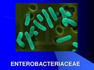
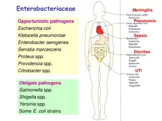
This is a large group of microorganisms that most of them live as a normal flora in the intestine (colon) of human and other animals and may be other parts of the body, and cause several enteric diseases as well as other diseases that involve the respiratory, genitourinary, the meninges and the skin. The classification and groups belong to this family will be studied during the theory lectures.
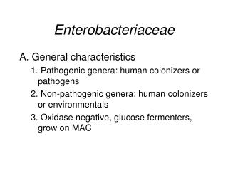
General characters: There are some general characters that are shared by all bacteria belong to this family, these are: 1- Gram negative non spore-forming rods. Some are motile, others are capsule producing. 2- Facultative anaerobes 3- Ferment glucose with or without gas 4- Not fastidious but can grow on ordinary media 5- Oxidase negative , catalase +ve
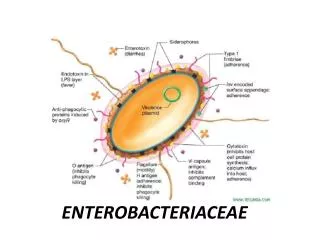
E. coli (Escherichia coli) Normal flora of human and animal GIT, human female genital tract. Under electron microscope
E. coli (Escherichia coli) • There are many STRAINS SEROTYPED according to O Ag, H Ag, K Ag. • Most strains are Motile, un-capsulated. • Produced dry colonies.
E. coli (Escherichia coli) infections DISEASES EXTRAINTESTINAL infections INTESTINAL infections
There are certain tests and media used for this group of bacteria for characterization, isolation • and differentiation between the different genera and also species in the same genus. Such media are: 1- MacConkey agar: (The main ingredients; bile salts, lactose and neutral red indicator) This is one of the important media used as a selective as well as a differential medium. As a selective because it contains bile salts that inhibits the growth of contaminated Gram positive cocci that may be found in the specimen, except for Strep.faecalis that normally live in the intestine. As a differential, because it contains lactose, therefore; the bacteria that ferment lactose will produce acid and change the color of the indicator to red, and the colony will appear red or pink in color, while the non lactose-fermentor will appear yellowish in color. (Exercise: try to observe the colors of different genera and species on this medium which indicates whether it is lactose-fermentor or non lactose-fermentor and write it down).
Dry, discreet pink colonies E.coli on MacConkey’s agar (pink colony due to lactose fermentation
Klebsiella on MacConkey’s agar (pink mucoid colony due to lactose fermentation
2- Eosin methylene blue (EMB) agar: (The main ingredients: Bile salts, eosin and methylene blue stains) This is also a selective and a differential medium. As a selective because it contains bile salts (see above), and as a differential; because there are certain bacteria that have the ability to combine these two stains and produce a violet greenish metallic sheen colonies, others do not posses this ability. (Excersice: observe this phenomenon and write down the bacteria that can and that can’t produce this phenomenon).
Non differential medium: Like Blood agar Enterobacteriaceae appears (large, gray, smooth)
3- Triple Sugar Iron (TSI) agar: (The main ingredients: 3 sugars (glucose, lactose and sucrose), iron salts, and phenol red indicator) This is one of the important differential media used for enterobacteriaceae. It detects whether the organism has the ability to ferment glucose only or all the 3 sugars, produce gas from fermentation or not, and also if the organism can split sulfur from protein and produce H2S or not. Glucose is present in only 0.1%, while the other 2 sugars are 1%. The medium is in the slant form, and the organism will be streaked on the surface and also stabbed by a needle to almost the bottom of the agar. During the first 6-8 hours all groups of bacteria will utilize and ferment glucose (by definition) with production of acid which will change the color of the indicator (phenol red) from pink to yellow in the whole tube.
Since glucose is only 0.1% therefore; after further incubation (18-24 hours) it will be soon exhausted, and if the bacteria capable of utilizing and fermenting the other two sugars, this means will continue to produce acid and the color will remain yellow at the slant and at the butt of the tube (slant: acidic, butt: acidic). On the other hand, if the bacteria are not capable of utilizing the other two sugars will switch to peptones present in the medium and splitting it to amino acids and then to ammonia as end product. Since the bacteria on the surface (slant) grow faster (aerobic) than those at the bottom of the tube (anaerobic), therefore glucose will be exhausted at the slant first and ammonia produced will re-change the color of the indicator to pink, therefore the tube will be read as (slant: alkaline (pink), butt: acidic (yellow)).
The gas produced (CO2) by fermentation will lead to cracking or even pushing the medium upwards, and this is a qualitative test for gas production. • Some species of bacteria has the ability to split sulfur from proteins and produce H2S as an end product; this will combine with iron ions forming FeS as a black precipitate coloring the medium. • N.B.: Kligler Iron Agar has the same principle of reaction except it contains two sugars ( glucose and lactose) instead of three. • (Exercise: try to observe and study all types of fermentations on this medium, and write down the organisms that produce them, and notice the gas production as well as H2S production and indicate the organisms that do so).
4- Sugar fermentation tests: (The main ingredients, broth medium with a single sugar, phenol red indicator, Durham tube) This test is used to detect the ability of an organism to ferment a specific sugar with or without production of gas. If the organism ferment the sugar the medium will look turbid, and the color of the indicator will be changed from pink to yellow. If there is gas produced from the bacteria at the bottom of the tube, it will be collected by the inverted Durham tube, and amount of the gas collected (which is quantitative) depend on the type of the organism, which could be used as a distinctive character.
lactose fermenting Non lactose fermenting
5- IMViC test : I : Indole production M: Methyl red test V: VogesProskeur test C: Citrate utilization This test is considered as the most practical test for identification and differentiation between thedifferent species belong to this family.
Indole production: ( the medium used is called peptone water which contains the amino acid tryptophan) Certain bacteria has the ability to split indole from tryptophan molecule. Tryptophan is an amino acid that can be oxidized by certain bacteria to form 3 major indole metabolites: Indole, Methyl indole (skatole) and indole acetic acid (indole acetate). Various enzymes are involved which are collectively called; tryptophanase: L-tryptophan Indole pyruvic acid Indole acetaldehyde Indole acetic acid Deamination Decarboxylation Indole (Skatole) Methyl indole
Indolesplit from the tryptophan molecule , could be detected by a reagent which is either Kovac’s reagent (or Erlichreagent) Kovac’sreagent: Pure amyl or isoamyl alcohol (or butyl alcohol)..….. 150 ml. P-dimethylaminobenzaldehy……………………….. 10.0 gm. Conc. HCl…………………………………………… 50.0 ml. Indolecombines with the aldehyde present in either Kovac’s or Ehrlich’s reagent to give a red colour( rosindole) floating on the alcohol layer, the negative will give a yellow colour. (Exercise: add the reagent to a 24 hour old culture on peptone water and observe the rings and write down the organisms that give +ve or –ve tests).
Laboratory Diagnosis Indole test
. Methyl red & VogesProskeur tests: (The medium used is called MR/VP medium) This test is used to determine the ability of an organism to continue fermenting the sugars and Producing stable acids despite the pH has reduced to a very low level (less than 4) by ,mostly, mixed acid type of fermentation. Methyl red indicator gives red color under pH of lower than 4, therefore addition of few drops of it to a 48-72 hours old culture will give a dark red color. On the other hand, other bacteria has no such ability, therefore as soon as the acids accumulated and the pH reduced they start to break down the initial fermentation products and also the pyruvic acid by decarboxylation and produce more neutral compounds, such as acetyl methyl carbinol (acetoin) which is a step prior to the 2,3 –butylene glycol product, leading to raise the pH to around 6. This will give methyl red test –ve and VogesProkeur test +ve. In other words, when methyl red test is +ve, VP will be negative and vice versa.
One molecule of acetoin is formed by the decarboxylation of 2 molecules of pyruvic acid; 2 Pyruvic acid Acetolactic acid + CO2 Acetoin + CO2 2,3-butylene glycol NADH NAD
V.P. reagent; 5% α – nephthol in absolute ethanol alcohol………. 6 drops 40% KOH + 0.3 gm. creatine ……………………… 2 drops α – nephthol is first added to which combines with acetoin to form diacetyl in the presence of air (O2) by shaking. Then KOH is added to facilitate the oxidation with the formation of a red-pinkish colour. (Exercise: try to do these tests, and write down the organisms that give +ve or –ve reactions).
Laboratory Diagnosis Methyl red test
Laboratory Diagnosis Voges-proskauer test
Citrate utilization test: (The medium used is called Simmon citrate medium with bromothymol blue indicator) The principle of the test depends on the ability of the organism to utilize citrate as the only carbon source in the medium, therefore; the organism will have no other choice either utilize it or it will mostly die. 4 Citrate 7acetate+5 CO2+Formate +Succinate
The medium is prepared as a slant in a tube and the organism is streaked on the surface and incubated for 24 hours. The organism that can utilize citrate as a carbon source has also the ability to utilize peptone as a nitrogen source leading to production of ammonia making the medium alkaline and lead to change the color of the indicator (bromothymol blue) from greenish to blue color giving the +ve test. On the other hand, if the organism has no such ability, it will not grow and no color change occurs. (Exercise; try to do the test and observe the color differences and write down the organisms that give +ve and –ve reactions)
Laboratory Diagnosis Citrate test
Laboratory Diagnosis 5) Motility test(at 37C°): E.coli causes inverted tree (Christmas tree) due to it’s motility (+ve), while Klebsiella doesn’t as it is not motile (-ve). Semisolid medium – motility test (inverted Christmas tree