Case studies in vascular surgery for MRCS
520 likes | 774 Vues
Case studies in vascular surgery for MRCS. Harj Rayt STR general and vascular surgery. What is an aneurysm?. What if its less dilated? What is arteriomegaly ?. Focal dilatation of a blood vessel to more than 1.5x the normal diameter.

Case studies in vascular surgery for MRCS
E N D
Presentation Transcript
Case studies in vascular surgery for MRCS Harj Rayt STR general and vascular surgery
What is an aneurysm? • What if its less dilated? • What is arteriomegaly? Focal dilatation of a blood vessel to more than 1.5x the normal diameter
What is the single most important factor in deciding when to operate on an AAA? Size Other criteria: operative fitness, co-morbidities UKSAT: 5.5cm requirement
When might you operate on a smaller aneurysm? Rupture Distal embolisation Back pain Growth rate of >1cm/yr
How would you manage a small (<5.5cm) AAA? USS surveillance
How would you image a patient with an aneurysm pre-operatively and what are you looking to identify? • Size • Shape • Relationship to renal arteries • Suitability for EVAR • Other aneurysms (iliac, thoracoabdominal) • Reasons not to operate (malignancy) • Reasons that might make surgery dificult (horsehoe kidney) CTA Angiogram MRA
Is screening for AAA effective? Several trials have shown this to be the case Benefits seen in Caucasian men over 65
Outline the principle steps in an elective AAA repair? • iv heparin • Prox and distal control • Sac opened, back bleeding from IMA and lumbars controlled • Graft chosen, cut to size and sutured with prolene • Top end tested before moving to bottom end • Sac, peritoneum and abdomen closed Transverse or midline incision Small bowel packed away Duodenum (4th part) mobilised Posterior peritoneum over aorta incised AAA neck, renal arteries and vein identfied Distal aorta identified (iliacs)
How do vascular and bowel anastomoses differ? • Vascular • Non-absorbable, monofilament – prolene • Continuous • Everting • Bowel • Absorbable • Continuous or interrupted • Inverting the mucosa
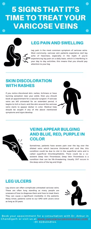
Detail how you would assess a patient with varicose veins? • Examination • Standing – assess both limbs • Skin examination • Groin for varix • LSV/SSV distribution • HH doppler for incompetence • Duplex USS • History • Duration & nature of symptoms • Interference with daily life • Family history • Occupation (standing) • PMH – DVT/ fracture • OCP
What is a saphena varix? • Dilatation of LSV just before the junction with the femoral vein. The valve here is incompetent • Examination • Lump in the groin • Empties on minimal pressure, refills on release • Fluid thrill on percussion
What operation would you perform for saphenofemoral incompetence? How? High tie, strip and avulsions
What is the recurrence rate? Variable 7-70% Approx 1/5 of all vv surgery is for recurrence
How would you distinguish between the LSV and femoral vein at surgery? Can be difficult LSV has tributaries joining Femoral vein receives only one branch (LSV) in the groin Need to trace the femoral vein prox and distally before any division of LSV
What are the tributaries of the LSV in the groin? Variable Superficial external pudendal Deep external pudendal Superficial circumflex iliac Superficial inferior epigastric
What are the indications for varicose vein surgery? Prevent complications from developing or progressing Varicose eczema, lipodermatosclerosis, ulceration Bleeding Superficial thrombophlebitis Pain Cosmesis (Not NHS)
What are the surface markings of the LSV? Drains the medial end of dorsal venous arch in foot Passes anterior to medial malleolus Ascends with saphenous nerve in superficial fascia in medial aspect of leg At knee, it lies a hand’s breath behind medial aspect of patella Passes along medial aspect of thigh, through saphenous opening in cribriform fascia to join femoral vein 2cm below and lateral to pubic tubercle
Define an embolus and give examples • ‘an abnormal mass of undissolved material carried from one place to another in the bloodstream’ • Thrombus • Tumour (RCC that invade the IVC) • Fat (long bone fractures) • Atheroma (ruptured plaques) • Gas • Amniotic fluid • Foreign body (catheter tips)
What are the 6 symptoms that define an acutely ischaemic limb? Pain Pallor Pulselessness Paraesthesia Paralysis Perishingly cold
A 63 y/o lady with AF develops a painful cold right leg. Give possible diagnoses and management plan • iv heparin • Embolectomy • GA/LA • Longitudinal skin incision • Expose and sling vessels (CFA, SFA, PFA) • Prox and distal control • Transverse arteriotomy • Fogarty catheter • Irrigate vessels with hep saline • Close primarily with prolene or with vein patch Femoral embolus related to AF
If there was residual thrombus in distal vessels, what adjunctive procedure could you perform? Thrombolysis tPA streptokinase
How would you manage this patient post-operatively? Resuscitation and monitoring of limb Identification and treatment of cardiac disease Physio for limb and chest Early mobilisation Continued anticoagulation
What would you do in a patient with a threatened limb, where angiography reveals a thrombosed popliteal aneurysm? Fem-pop bypass Grafts: dacron, PTFE, reversed vein graft
What are the indications for lower limb amputation? Unsalvagable limb Septic limb Painful ischaemic limb with no viable reconstruction Malignancy Trauma Infections - polio
What level should the amputation be performed? High enough to remove all dead tissue and allow healthy tissues to heal Think about rehab
Describe the principle steps in a BKA Mark for long posterior flap Skin flaps raised and muscles divided Ligate LSV Elevate tibialperiosteum and divide 1cm above skin incision Fibula divided 1cm higher Post flap fashioned and muscles divided at an angle Remaining vessels ligated, sciatic nerve divided high Haemostasis, drain Closure with vicryl to fascia and prolene to skin
What are the contraindications to a BKA? Need for higher amputation Fixed flexion deformity of the knee Inability to leave a long tibial stump (min 7.5cm) Insufficient tissue for adequate healing
What are the complications of a BKA? Haematoma Infection Poor healing Stump breakdown, need for revision Phantom limb pain
What are the indications for the elective repair of an AAA? • UKSAT – repair of AAA 4-5.5cm proved no survival benefit when compared to patients who had serial USS • Repair if: • Symptomatic • >5.5cm in size • Growth of >1cm/annum
What pre-operative measures should be taken? • Assess for co-morbidity: lungs, renals, cardiac • Liaise with anaesthetist • Investigations include • Bloods inc XM • ABG • PFT • CXR • ECG • ECHO • ETT • Image AAA
What are the complications of AAA repair? Immediate – haemorrhage, trash foot, distal limb arterial thrombus Early – spinal cord ischaemia, mesenteric ischaemia, ARF, MI, CVA Late – false aneurysm formation, graft infection, aortocaval/duodenal fistula, impotence
Talk about EVAR… Criteria How to do Complications
What are the surface markings of the abdominal aorta? Begins at aortic hiatus through diaphragm at T12 Ends at L4 with bifurcation into iliacs 3 unpaired branches – T12 coeliac axis, L1 SMA, L3 IMA
What are the indications for CEA? • Symptomatic patients with carotid artery territory symptoms • Amaurosis fugax • Hemiplegia • Dysphasia • evidence of cartoid artery stenosis between 70-99%
What is the difference between carotid artery territory symptoms and vertebro-basillar symptoms? • Carotid artery territory • Hemianopia, contralateral hemiparesis, dysphasia • Vertebro-basillar • Vertigo, diplopia, dysphagia, dysphonia, nausea, vomiting, dizziness, ataxia
What pre-op imaging should be taken? DUSS – assess anatomy, quantify degree of stenosis, look for occlusion, assess contralateral side. Can be combined with TCD Angio – carries risk of CVA CT/MRA – useful for assessing other lesions, ie subclavian stenosis CT brain
Are you aware of any trials that support CEA? North American Symptomatic Carotid Endarterectomy Trial (NASCET) CEA beneficial to patients with a symptomatic carotid stenosis of 70-99% Follow up: 2 yr risk of ipsilateral CVA reduced from 26% to 9% (P<0.001) NNT to prevent 1 CVA in 2 yrs = 6 European Carotid Surgery Trial (ECST) CEA reduced absolute risk of CVA and death over 3 yrs from 21.9% to 12.3% (P<0.01)
During the procedure, how can you distinguish the ECA from ICA? Origin of ECA is anteromedial to origin of ICA ICA has no branches outside the skull
Which nerves are at risk during CEA? Vagus Hypoglossal Glossopharyngeal Ansacervicalis