Download
1 / 24
280 likes | 599 Vues
Brain Imaging Core: MRI of Structure, Microstructure, Metabolites, and Animals. Andrew L. Alexander, Ph.D. alalexander2@wisc.edu Departments of Medical Physics & Psychiatry Waisman Laboratory for Brain Imaging and Behavior University of Wisconsin - Madison. Magnetic Resonance Imaging (MRI).

E N D
Brain Imaging Core: MRI of Structure, Microstructure, Metabolites, and Animals Andrew L. Alexander, Ph.D. alalexander2@wisc.edu Departments of Medical Physics & Psychiatry Waisman Laboratory for Brain Imaging and Behavior University of Wisconsin - Madison
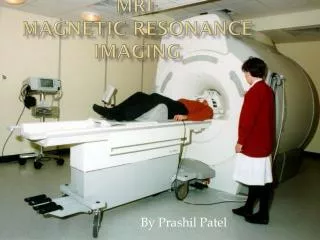
Magnetic Resonance Imaging (MRI) Large magnet • 3 Tesla! • 10,000 times Earth’s magnetic field • Superconducting (magnet is always on) • Cooled with liquid helium (-265 °C) • 11,000 kg. • ~200km of wire Rasmus M. Birn Send radio waves into body (no X-rays)
Structural MRI of the Brain T2-weighted T1-weighted
Morphometry – Size and Shape of Brain Structures AJNR 2001 Moo Chung
Cortical Thickness - Freesurfer Greg Kirk
Diffusion Tensor Imaging • Non-invasive imaging technique for estimating the diffusion properties of water in biological tissues • Highly sensitive to differences in white matter microstructure • White matter connectivity properties influence microstructure and diffusion Anisotropy (‘White Matter’) T2W Structure Orientation of WhiteMatter Total Diffusion (1/Density) Axonal Density Diffusion Myelin Diffusion
Diffusion probes tissue microstructure ADC = Apparent Diffusion Coefficient More barriers Fewer barriers ADC low ADC high
The Diffusion Tensor is a Matrix z z e2, l2 e1, l1 y y e3, l3 x x
z y z x y x Diffusion Tensors z y x
Study Specific DTI Templates N=270; Adluru et al. submitted Rhesus NH Primate Template Human Template
Rhesus Tractography Human Tractography NageshAdluru, Do Tromp
Tracula • Automated Global Tractography Method - Freesurfer Greg Kirk; From A. Yendiki 2011 TRACULA
Framework of modeling human connectome using dMRI [Zalesky et. al., 2010]
Structural Network Connectivity Mapping Chung 2009
Degree of Connectivity Control Autism Chung 2009
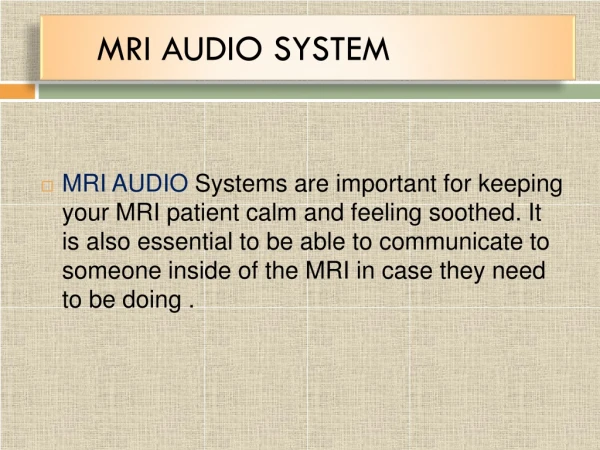
MR Spectroscopy MR spectroscopy provides chemical information. Measurement can be localized to a specific brain region. Like other MR imaging, MR spectroscopy is non-invasive. Can do (coarse) spectroscopic imaging 1H MR spectroscopy can be added as one or more additional scans in an MR imaging protocol. Data is collected from a single voxel (typically 8mL voxel, 7 minute acquisition). Offline data processing with LC Model determines the concentrations of about 16 metabolites. Lisa Angelos
Fitting and Metabolites Lisa Angelos NAA mI Cho Cre
Amygdala Spectroscopy • Protocol to collect data from the amygdala • Oblique localizer scans to visualize anatomy • Saturation bands help isolate amygdala signal • 7-minute acquisition, LC Model processing • Corrections for tissue content and voxel edge location • Reproducibility study • Five major metabolites (NAA, Cre, Cho, mI, Glx) measured with better than 20% accuracy • In a healthy adult sample, NAA and Cre individual differences detected with ICC = 0.6. Lisa Angelos
Spectral Editing (MEGA-PRESS) Every other acquisition has freq range suppressed GABA refocusing Difference shows GABA refocusing (Cre cancels). Every other acquisition is “normal” GABA minimized (TE=68) TE=35 spectrum Red line: GABA + baseline Lisa Angelos
Lisa Angelos Phenylalanine in PKU water peak removed “downfield” from water “upfield” from water Small phe peak overlaps large tryptophan peak
Other Details • Image Processing / Statistics • Canned Software Packages • Custom Software Tools • Advice and Interpretation • Atlasing • Studies in humans, NHP, dogs, cats • Ex vivo possible
Other Methods • Myelin Characterization: Magnetization Transfer, Myelin Water Fraction Mapping (Relaxometry) • Iron Mapping (T2, T2*) • High b-value diffusion imaging / complex white matter characterization • Image guided surgery / drug delivery • Contrast agents • Scanning in Children – Infants to Adults
Pre-Clinical MRI Lab Varian 4.7T imaging system -scan small animals up to 600 grams - in-plane resolution on the order of 50 microns. It has broadband capability allowing us to scan a variety of nuclei including H1, P31, F19 and C13. This system has the ability to perform a wide array of MRI sequences including T1 and T2 weighting, T1, T2 and T2* mapping, functional MRI (EPI), diffusion and diffusion tensor imaging, localized spectroscopy (STEAM and PRESS) as well as chemical-shift imaging, and perfusion imaging with Gadolinium based contrast agents. This allows for visualization and quantification of a variety of moieties and processes including metabolites, anatomical structures, tumor morphology, blood flow/vessels, fiber pathways, drug effects, brain activity, and heart motion. Our system is equipped with a variety of surface coils and dual tune volume coils. In addition, our lab system includes a Carbon-13 Hyperpolarizer. High signal to noise and high resolution are two essential factors for successful molecular imaging. Normally, MR provides poor signal to noise especially when attempting to image molecules containing low natural abundance nuclei, such as carbon-13. Recently, a new commercially available dynamic nuclear polarizer (DNP), the Hypersense system by Oxford instruments, has been acquired that is able to increase the signal to noise available for carbon-13 by over 10 000 times. This enables the rapid acquisition of images from injected substrates such as carbon-13 labeled pyruvate and any compounds that may be produced via its in vivo metabolism. The system works by freezing a solution of a carbon-13 enriched substance mixed with a paramagnetic compound to just above 1 kelvin inside a 3 tesla magnet. The mixture is microwaved (to transfer polarization from electrons to the carbon-13 nuclei) and, after a period of time, heated quickly using super-heated water to enable the hyperpolarized material to be extracted for injection. This system provides many opportunities to study perfusion, angiography and metabolism in a variety of in vivo models. CONTACT INFORMATION: if you are interested in our services, wish to tour our facility, or arrange a meeting discuss how we can assist your research, please contact Beth Rauch at 608-265-1109 or brauch@wisc.edu
Pre-Clinical MRI Lab Scheduling and Fee Information: All users must first schedule a planning session to discuss how our services can best meet your needs. Please contact Beth Rauch at brauch@wisc.edu or 608-263-1109 to do so. Fee Structure: *the preliminary scans for start-up for new projects are free of charge. UWCCC Members: $220/hour Non UWCCC Members: $375/hour Commercial Users (non UW): $556/hour Anesthesia & Supply Charge: $5/scan Data Analysis: $30/hour Technical Development Time: $40/hour Contact us for a free consultation at brauch@wisc.edu or 608-263-1109. • Potential Uses of the System: • Ex-vivo and In-vivo Imaging • Molecular imaging • Track stem cells in vivo • Macrophage infiltration • Pin-point brain ischemia • Tracking neural pathways • Image Anatomy • Transgenic Animals • Oncology • Phenotyping • Atherosclerosis • Neuological disorders The Carbone Cancer Research Imaging Center at the University of Wisconsin features a Varian 4.7T horizontal bore imaging/spectroscopy system. This system provides the capability to scan samples up to 600 grams in size, or with a diameter of 72 mm or less, with an in-plane resolution in the order of 50 microns. It is also equipped with an anesthesia system and physiologic monitoring that allows for image gating. MRI is totally non-invasive (there is no ionizing radiation) and scans can be repeated multiple times to track disease progression or treatment effectiveness. This system is a shared resource for the UW system and is the only one of it’s kind on campus. www.medphysics.wisc.edu/preclinmri.html