Brain imaging
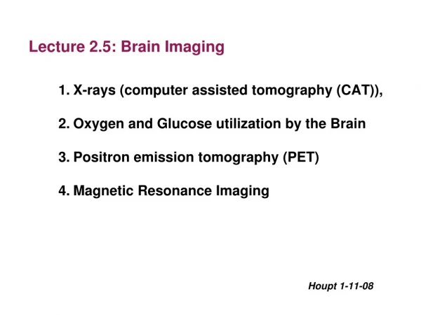
Brain imaging. Take a look inside!. NEUROIMAGING. Neuroimaging is the capture of detailed images of the living intact brain as people engage in different mental processes or make behavioural responses. The various techniques can be divided into 2 general categories:


Brain imaging
E N D
Presentation Transcript
Brain imaging Take a look inside!
NEUROIMAGING • Neuroimagingis the capture of detailed images of the living intact brain as people engage in different mental processes or make behavioural responses. • The various techniques can be divided into 2 general categories: • Structural – scanning techniques that show brain structure and anatomy (CT, standard MRI). Typically show cross sections of brain. • Funtional– scanning techniques that provide views of a particular aspect/s of brain function by showing the brain “at work”. They also provide information about brain structure. (PET, SPECT and fMRI)
Electrical Stimulation - ESB • Involves using an electrode to deliver a precisely regulated electric current to the brain, thereby stimulating a specific area of the brain • If electrical stimulation of a specific brain area initiates a behavioural response, then that specific area of the brain controls or is involved in that response • Extremely invasive – usually only conducted when other surgery is already necessary • It may stimulate a response or disrupt brain functioning so impairing a response.
PENFIELD’S Studies • Early Direct Brain Stimulation studies conducted by US neurosurgeon Wilder Penfield in the 1940s to 1960s mapped the functions of various parts of the cerebral cortex with numbered tags. • This was done as part of surgery to treat epilepsy. Patient was conscious during the procedure where the area of the cortex thought to specifically be the source of the epileptic seizures for last resort patients who experienced life-threatening seizures.
Penfield’s studies • Patients’ brain was stimulated with an electrode and then asked to report their experiences. • E.g. Occipital lobe stimulation generated patients reporting seeing flickering lights, spots, colours, stars and other visual images. • Auditory cortex stimulation – patients reported “hearing” doorbells, engines and a range of sounds. • Primary somatosensory cortex stimulation – patients reported “feeling” a tingling sensation in their cheek, hand, leg and other body parts. • Primary motor cortex stimulation – patients responded by moving specific body parts. This led to mapping the motor cortex.
Penfield’s mapping of cortex • Some cortex sites generated “experiential responses” such as patient vividly recalling past events not considered for years. • Data from hundreds of patients was pooled to map cortical areas and their functions. The map has since been confirmed by other researchers. • Ojemann and Mateer (1979) – investigated direct stimulation on specific language areas of brain – bilingual patient showed stimulation of different brain areas interfered with either English or Spanish use but not both.
Deep Brain Stimulation • Electrodes have also been used for therapeutic purposes – known as “deep brain stimulation”. • Electrodes planted deep in the brain of patients with severe and resistant depression showed striking and sustained remission of depression. The stimulation reduced activity in a brain area believed to be overactive in depression.
Transcranial Magnetic Stimulation TMS • Delivers a magnetic field pulse through the skull stimulating the neurons closest to the point of stimulation • Temporarily activates or disrupts normal activity of neurons in specific cerebral cortex area. • Only effects neurons to a depth of 2 – 3 centimeters\ • Good for establishing how different brain regions control different functions • Allows for simulated brain damage • Long term effects of repeated exposure are unclear • Side effects can include localised pain or headache Non invasive, no substances or anaesthetic required
Transcranial Magnetic Stimulation • The magnetic field induces a harmless electric current in time varying charges (“pulses”). • Person is fully awake and alert. • Single pulse TMS (‘non-repetitive TMS”) is when the procedure involves only a single pulse. • Repetitive TMS (“rTMS”) is when procedures involve repeated but not necessarily rapid delivery of a pulse. • A small copper electromagnetic coil that is enclosed in plastic and placed next to the scalp. Electric current is sent through the coil that induces a magnetic field around the coil and creates the pulse.
Transcranial Magnetic Stimulation • The pulse activates the neurons and they send a burst of neural impulses to adjacent neurons, activating them, which in turn activates other adjacent neurons. • Placed over occipital lobe – flashes of light detected • Placed over motor cortex – brief muscle twitch somewhere on body depending on which part of motor cortex • Pulse does not directly affect whole brain. • TMS used to study specific areas of cerebral cortex. • TMS can be used for mapping brain areas.
TMS used for.. • Patients who have had a stroke, suffered a brain injury, or spinal injury – to help pinpoint specific areas of cortical brain damage and to track patient recovery. • Can be used for pre-surgical assessment of patients with brain tumours and neural diseases, Parkinson’s, motor neuron disease and MS. • LIMITATIONS • Only affects the part of the brain directly below the skull
rTMS • When rTMS is used, the consecutive pulses cause the neurons to lose their ability to fire, resulting in suppression of their activity and consequently brain activity in the stimulated area. • E.g. Coil held over Broca’s area, person is unable to speak fluently while current is on but will resume when current stops. • rTMS allows researchers to temporarily create brain “malfunction”, simulating brain damage, which permits them to perform experiments with human participants previously impossible. E.g. Cause and effect studies • rTSM produces effects that last longer than single pulse TMS.
Using rTMS to treat depression • 67 Israelis – independent groups using double blind procedure. • One group received rTMS to left frontal lobe cortex area daily for 30 mins. for 2 weeks. • Other group received placebo treatment (without TMS). • At the end of 2 weeks, half of stimulated patients showed at least 50% improvement in scores on a scale used to measure depression compared with 25% in placebo group. • Not used currently widely yet to treat depression as it seems that rTMS is capable of changing the activity in a brain area, even beyond the duration of the rTMS application itself.
Value and Limitations of Direct Brain Stimulation • Advanced our understanding of role of brain in mental processes and behaviours. • Able to identify locations and functions of numerous brain structures and areas, as well as hemispheric specialisation. • Extremely invasive. • Difficulty in generalising results from research with epileptic patients to normally functioning brains. • Future potential use of electrode implants to control chronic brain illnesses or disorders.
Value and limitations of TMS • TMS is non-invasive. • No sedation or anaesthetic required. • No X-rays or radioactive substances used. • Can’t be used with patients who have metal or implanted medical devices in their body. • Can’t be used with patients who have seizures. • Long term effects of repeated stimulation not yet established – especially for fast and high intensity pulses. • Scalp pain, mild headache – side effects of rTMS • Can induce seizures • Can only go as deep in the brain as the cerebral cortex
Computerised axial tomography (CAT) Scan • A computer enhanced X-ray of a slice (cross-section) of the brain created from X-rays taken from different angles. • CT is extremely useful for identifying the precise location and extent of damage to or abnormalities in various brain structures or areas. A CT scan can reveal the effects of strokes, tumors, injuries and other brain disorders • It does not provide information about the activity of the brain
Computerised axial tomography (CAT) Scan • Involves moving an X-ray source in an arc around the head while a computer compiles different “snapshots” of the brain area. • Can only be performed by a radiologist. • Patient given an injection of a substance called “contrast” based on iodine into vein of their arm or hand. “Contrast” is not radioactive, rarely has serious side effects, but mild reactions can be experienced by approx. 39% of people. • The “contrast” highlights the blood vessels which assists in interpreting the CT images.
CAT scan procedure • Patient lies still on a CT scanner bed. • Horizontal cross-sectional image produced is only a few millimetres thick. • The image produced is known as a “CT scan” or “CAT scan”. • Provides far greater clarity and detail than a conventional X-ray. • Non-invasive procedure. • Radiation dosage during X-ray procedure is considered relatively harmless.
Value of CT scans • Extremely useful for spotting and identifying the precise location and extent of damage to or abnormalities in various brain structures or areas. • Used to identify abnormalities in brain structures among people with mental illnesses (e.g. Schizophrenia, depression) • Has enabled observation of physical changes or differences in the brains of patients with brain-related disorders such as Parkinsons and Alzheimers.
Limitations of CT scans • Shows only brain structure or anatomy. • Not information about brain functioning or activity.
POSITRON EMISSION TOMOGRAPHY (PET) • Prior to the scan being taken, the person is given a sugar-like substance that contains a harmless radioactive element. When this substance enters the bloodstream it travels to the brain. As particular parts of the brain are activated, the substance emits radiation which is detected the PET scanner. • Great for examining brain function when performing different tasks • As it involves radioactive element regular use is to be avoided
PET cont... • Uses a colour code to indicate levels of activity – provides images of the “working brain” by tracking blood flow around the brain. Violet, blue, green, yellow, red • Eg if a person is listening to someone talking during the PET procedure, the areas involved with speech production would be activated and highlighted in red and yellow – ieWernike’sarea • Assumes increased blood flow indicates increased neuronal activity (and vice versa). • Blood flow is tracked by measuring the use of glucose (sugar) by neurons in the brain area that is active. • The type of tasks that can be given to a participant is limited to the size of the chamber, so PET studies are often relatively simple.
PET cont’d... • Prior to PET, a harmless radioactive substance is injected into the blood vessels or taken orally. As it enters the bloodstream it travels to the brain. • When a particular area of the brain is activated during mental or behavioural processes the radioactive substance serves as a tracer that can be followed and emits radioactive signals.
Advantages & Limitations ADVANTAGES: Highly effective in identifying brain abnormalities Eg Brain tumours usually very active Brain functioning & changes in brain can be identified LIMITATIONS: Researchers cannot determine whether an active brain area is actually involved in the mental process or behaviour under investigation. Radioactive dye decays rapidly – can only be used for short tasks 40sec interval between scans, each scan lasts only 30 secs.
Single photon emission computed tomography (SPECT) • Similar to PET but differs in certain technical features • Radioactive tracer lasts longer • Uses scanner to record data • Computer uses data to construct 2 or 3 dimensional images of brain active regions. • Not as detailed as PET – SPECT images have a lower resolution, so not as clear • Can be combined with CT images to increase resolution and more accurately pinpoint location of brain structure or abnormality • Much cheaper than PET
Advantages and Limitations • Advantages: indicates function, rather than being purely anatomical, tracer longer lasting so longer tasks can be performed. • Accurately pinpoints location of abnormality when combined with a CAT scan • Much less expensive than PET • Disadvantages: not as clear or detailed as PET images
Magnetic resonance imaging (MRI) • MRI uses a similar technique to the CAT scan, but instead of using an X-ray, harmless radio frequencies are used to vibrate atoms in the neurons of the brain to produce image • The amount of vibration is detected and analysed by a computer • MRI can be used to detect and display extremely small changes in the brain. For example, MRI can more clearly distinguish between braincells that are cancerous and those that are noncancerous also tissue degeneration & blood clots/leaks that may indicate a stroke. • shows only brain structure not function
Advantages & Limitations • ADVANTAGES • Enables more precision in the study of live brains • Non-invasive and harmless • More detailed than CT, almost photographic in quality • Does not use radio-active substances DISADVANTAGES • Cannot be used with people who have internal metal devices eg pace makers or pins in bones • Shows only structure (anatomy) not function
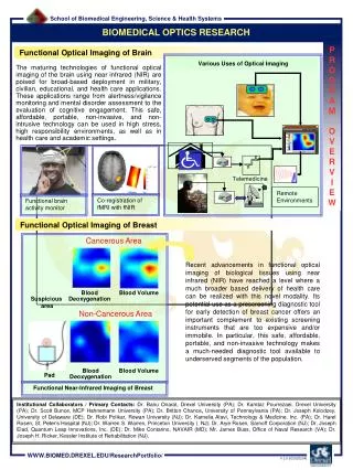
Functional magnetic resonance imaging (fMRI) • The technique is based on the standard MRI, and measures subtle changes in blood–oxygen levels in the functioning brain. • When an area of the brain is active, there is increased blood flow to that area, as more oxygen is required by the active, functioning neurons • Colour variations indicate neural activity • 3D image available from scans
Advantages & Limitations ADVANTAGES • Takes numerous images in rapid succession, therefore can detect changes from moment to moment. • More detailed and precise than all other scans • 3D virtual reality imaging possible • Detailed images of the functioning brain while performing tasks • Colour coding makes it easy to interpret. DISADVANTAGES • Very expensive • Limited access to fMRI
Ethics in research No, you cant just mash peoples brains and call it research!
Lobotomy • Steel rod inserted through the eye socket • Steel rod tapped through with a hammer then moved laterally to destroy brain tissue • Forward area of Frontal lobes destroyed • Used to treat everything from depression to schizophrenia and ADHD
Ethics – do the right thing! • Standards that guide individuals to identify good, desirable, or acceptable conduct • Australian Psychological society has a code of ethics • Also a National statement on ethical conduct on human research (2007)
Ethics – do the right thing! • Integrity – did this procedure/research genuinely show the potential to advance our understanding of the brain? • Respect for persons – were the welfare, rights, cultural heritage etc of the participants respected? • Beneficence – did the benefits of this procedure outweigh the potential risks? • Justification – is the use of this particular participant justified, do they represent the norm, will others benefit from the generalisation of these results.
Participants Rights • Informed consent • Withdrawal rights • Deception? • Debriefing • Confidentiality Do not talk about fight club!
Ethics committees – HREC (Human research Ethics Committee) • Ensure the study designed to meet national guidelines • Ensure researchers adequately qualified • Monitor progress • Handle complaints • Ensure accountability of researcher
Why use animals in research? • Some studies cannot be conducted on humans due to the risk of psychological or physical harm • Bodily systems and nervous system very similar in humans and many animals • Practical advantages – aging, breeding, • Animal behaviour can be controlled easily, environment can be controlled, thus extraneous variables are easily controlled • Expectations are not a factor in most animal research
Problems with using animals? • Generalising from animals to humans is not always possible • Harm to the animal; is it necessary? • All research covered by the Australian code of practice for the use of animals for scientific purposes • Justification – potential benefits must outweigh harm • Pain/distress only if no other alternative • Surgery must be with anaesthetic • Termination must be quick and painless
Things to do • Learning activities 4.24, 4.26, 4.27, 4.30 • True/False Quiz – pg 264 • Chapter 4 Test – pg 265 • StudyOn – Mind, Brain & Body, #s 5, 6, 7, 8