Brain imaging
Brain imaging. Take a look inside!. DIRECT BRAIN STIMULATION. Electrical Stimulation - ESB. Involves using an electrode to deliver a precisely regulated electric current to the brain, thereby stimulating a specific area of the brain If electrical stimulation of a specific


Brain imaging
E N D
Presentation Transcript
Brain imaging Take a look inside!
DIRECT BRAIN STIMULATION Electrical Stimulation - ESB • Involves using an electrode to deliver a precisely regulated electric current to the brain, thereby stimulating a specific area of the brain • If electrical stimulation of a specific brain area initiates a behavioural response, then that specific area of the brain controls or is involved in that response • DISADVANTAGE: Extremely invasive – usually only conducted when other surgery is already necessary
DIRECT BRAIN STIMULATION Transcranial Magnetic Stimulation TMS • Delivers a magnetic field pulse through the skull stimulating the neurons closest to the point of entry • Good for establishing how different brain regions control different functions • sTMS: Delivers a single magnetic pulse which activates neurons, thus activating activity of a brain region. • rTMS: Delivers multiple magnetic pulses that cause neurons to lose their ability to fire, thus suppressing activity • DISADVANTAGE: Side effects can include localised pain or headache
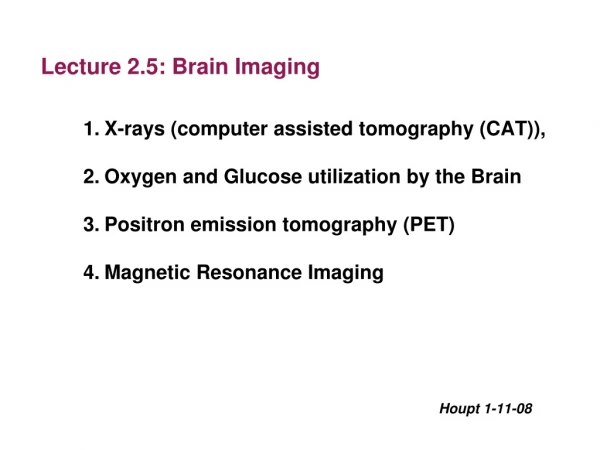
STRUCTURAL NEUROIMAGING Computerised axial tomography (CAT) Scan • A computer enhanced X-ray of a slice (cross-section) of the brain created from X-rays taken from different angles. • CT is extremely useful for identifying the precise location and extent of damage to or abnormalities in various brain structures or areas. A CT scan can reveal the effects of strokes, tumors, injuries and other brain disorders. • DISADVANTAGE: It does not provide functional information about the brain
STRUCTURAL NEUROIMAGING Magnetic resonance imaging (MRI) • MRI uses a similar technique to the CAT scan, but instead of using an X-ray, harmless radio frequencies are used to vibrate atoms in the neurons of the brain • The amount of vibration is detected and analysed by a computer • MRI shoes more clear images than CT • DISADVANTAGE: Shows only brain structure not function, can’t be used by people who have pacemakers.
FUNCTIONAL NEUROIMAGING POSITRON EMISSION TOMOGRAPHY (PET) • Prior to the scan being taken, the person is given a sugar-like substance that contains a harmless radioactive element. When this substance enters the bloodstream it travels to the brain. As particular parts of the brain are activated, the substance emits radiation which is detected the PET scanner. • Great for examining brain function when performing different tasks • DISADVANTAGE:As it involves radioactive element regular use is to be avoided
FUNCTIONAL NEUROIMAGING Single photon emission computed tomography (SPECT) • Similar to PET • Radioactive tracer lasts longer • Can be combined with CT images to increase resolution • Much cheaper than PET • DISADVANTAGE: Not as detailed as PET
FUNCTIONAL NEUROIMAGING Functional magnetic resonance imaging (fMRI) • The technique is based on the standard MRI, and measures subtle changes in blood–oxygen levels in the functioning brain. When an area of the brain is active, there is increased blood flow to that area, as more oxygen is required by the active, functioning neurons. • DISADVANTAGE: No cause-effect relationship like TMS
Ethics in research No, you cant just smash peoples brains and call it research!
Lobotomy • Steel rod inserted through the eye socket and tapped through with a hammer then moved laterally to destroy brain tissue • Forward area of Frontal lobes destroyed • Used to treat everything from depression to schizophrenia and ADHD
Ethics – do the right thing! • Standards that guide individuals to identify good, desirable, or acceptable conduct • Australian Psychological society has a code of ethics • Also a National statement on ethical conduct on human research (2007)
Ethical principles in brain research • Integrity – did this procedure/research genuinely show the potential to advance our understanding of the brain? • Respect for persons – were the welfare, rights, cultural heritage etc of the participants respected? • Beneficence – did the benefits of this procedure outweigh the potential risks? • Justice – is the use of this particular participant justified, do they represent the norm, will others benefit from the generalisation of these results.
Scenario • A researcher plans to conduct an EEG study on patterns of electrical activity in the brain that are associated with tasks involving thinking (eg. Solving arithmetic and word problems) as compared with voluntary movements (eg. Wiggling toes, rotating the knee, walking on a treadmill). The researcher proposes to compare responses of two groups of participants: people with brain damage to the left hemisphere and people with brain damage to the right hemisphere. All the brain damaged participants have been used by the researcher as participants in two previous brain studies and provide the advantage of being readily accessible because they reside in special- care accommodation close to the university where the researchers lecture on neuropsychology. The researcher does not expect any difficulties in obtaining informed consent. The researchers expects that the study will advance understanding of hemispheric functioning because the EEG has newly developed electrodes that are renowned for their exceptional sensitivity. The study also provides an opportunity to use the refined EEG technology and possibly refine the technique even further.