Pneumonia
Pneumonia. Community acquired pneumonia (CAP). Objectives. Discuss the epidemiology and pathophysiology of pneumonia and CAP Explain the different classifications of pneumonia Recognize clinical presentations associated with CAP Discuss the diagnosis and treatment of CAP

Pneumonia
E N D
Presentation Transcript
Pneumonia Community acquired pneumonia (CAP)
Objectives • Discuss the epidemiology and pathophysiology of pneumonia and CAP • Explain the different classifications of pneumonia • Recognize clinical presentations associated with CAP • Discuss the diagnosis and treatment of CAP • Identify common etiological agents causing CAP and discuss their laboratory work up • Discuss virulence factors and prevention of Streptococcus pneumoniae
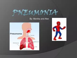
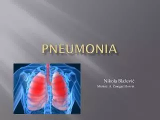
Definition • Pneumonia is an infection that leads to inflammation of the parenchyma of the lung (the alveoli) (consolidation and exudation) • It may present as acute, fulminant clinical disease or as a chronic disease with a more prolonged course
Epidemiology • Overall the rate of CAP 5-6 cases per 1000 persons per year • Mortality 23% • High, especially in old people • Almost 1 million annual episodes of CAP in adults > 65 yrs in the US Risk factors • Age < 2 yrs, > 65 yrs • Alcoholism • Smoking • Asthma and COPD • Aspiration • Dementia • Prior influenza • HIV • Immunosuppression • Institutionalization • Recent hotel : Legionella • Travel, pets, occupational exposures-birds (C. psittaci)
Etiological agents Infectious: • Bacterial • Fungal • Viral • Parasitic Non-infectious like: • Chemical • Allergen related
Pathogenesis Two factors involved in the formation of pneumonia • Pathogens • Host defenses.
Defense mechanism of respiratory tract • Filtration and deposition of environmental pathogens in the upper airways • Cough reflux • Mucociliary clearance • Alveolar macrophages • Humoral and cellular immunity • Oxidative metabolism of neutrophils
Pathophysiology • Inhalation or aspiration of pulmonary pathogenic organisms into a lung segment or lobe. • Results from secondary bacteraemia from a distant source, such as Escherichia coli urinary tract infection and/or bacteraemia (less commonly). • Aspiration of oropharyngeal contents (multiple pathogens).
Classification • Pneumonia classified according to: • Pathogen • Bacterial • Typical • Atypical • Viral • Fungal • Parasite • Anatomy • Acquired environment
Classification by anatomy 1. Lobar: entire lobe 2. Lobular: (bronchopneumonia). 3. Interstitial
Classification by acquired environment • Community acquired pneumonia(CAP) • Hospital acquired pneumonia(HAP) • Nursing home acquired pneumonia (NHAP)
CAP-fever+ productive cough + infiltrate • CAP : pneumonia acquired outside of hospitals or extended-care facilities Typical Atypical • Strept. pneumoniae • (lobar pneumonia) • Haemophilusinfluenzae • Moraxella catarrhalis • S. aureus • Gram-negative organisms • Atypical: not detectable on gram stain; won’t grow on standard media • Mycoplasma pneumoniae • Chlamydia pneumoniae • Legionella pneumophila
Community acquired pneumonia • Strep pneumonia 48% • Viral 23% • Atypical orgs (MP,LG,CP) 22% • Haemophilus influenza 7% • Moraxella catharralis 2% • Staph aureus 1.5% • Gram –ive orgs 1.4% • Anaerobes
Typical pneumoniaClinical manifestation • The onset is acute • Prior viral upper respiratory infection • Respiratory symptoms • Fever • Shaking chills • Cough with sputum production (rusty-sputum) • Chest pain- or pleurisy • Shortness of breath
Diagnosis • Clinical • History & physical • X-ray examination • Laboratory • CBC- leukocytosis • Sputum • Gram stain- 15% • Culture • Blood culture-5-14% • Pleural effusion gram + culture Pneumococcal pneumonia
Streptococcus pneumoniae • Gram positive diplococci • Alpha hemolytic streptococci • Catalase negative • Normal flora of upper respiratory tract in 20-40% of people • Causes: • Resp infections • pneumonia, sinusitis, otitis, • Non resp infections • bacteremia, meningitis
Streptococcus pneumoniae • Virulence factors: • Capsule • More than 90 capsular types • Pneumolysin • Autolysin • Neuraminidase • Prevention: vaccination
Streptococcus pneumoniae • Sensitive to Optochin • Lysed by bile (bile soluble)
Atypical pneumonia Approximately 15% of all CAP Not detectable on gram stain Won’t grow on standard media Some don’t have a bacterial cell wall Don’t respond to β-lactams • Chlamydia pneumonia • Mycoplasma pneumonia • Legionellaspp • Psittacosis (Chlamydia psittaci) • Q fever (Coxiellaburnettii)
Atypical pneumonia Symptoms Signs Minimal Low grade fever Few crackles Rhonchi • Insidious onset • Mild to severe • Headache • Malaise • Fever • Dry cough • Arthralgia / myalgia
Diagnosis & Treatment • Treatment: • Macrolide • Quinolones • Tetracycline • B lactams have no activity • Treat for 10-14 days • Diagnosis: • X-ray • CBC • Mild elevation WBC • U&Es • Low serum Na (Legionalla) • LFTs • ↑ ALT • ↑ AlkPhos • Sputum Culture on special media (BCYE) for Legionella • Urine antigen for Legionella • Serology for detecting antibodies • DNA detection
Mycoplasma pneumonia May be associated with extra pulmonary findings: skin rash, hemolysis, myocarditis, pancreatitis, encephalitis Diagnosis: Serology NAAT Culture can be done but requires special media and slow grower (weeks) • Eaton’s agent (1944) • No cell wall • Common • Rare in children and in > 65 • People younger than 40. • Crowded places like schools, homeless shelters, prisons. • Can cause URT symptoms • Usually mild and responds well to antibiotics. • Can be very serious
Chlamydia pneumonia • Obligate intracellular organism • 50% of adults sero-positive • Mild disease • Sub clinical infections common • 5-10% of community acquired pneumonia • Diagnosis: • Serology • NAAT
Psittacosis • Chlamydia psittaci • Exposure to birds • Bird owners, pet shop employees, vets • Parrots, pigeons and poultry • Birds often asymptomatic
Q fever (Coxiellaburnetti) • Exposure to farm animals mainly sheep • Spread by inhalation of infected animal birth products • Pneumonia is acute form of infection • Diagnosis: serology
Legionella pneumophila Legionnaire's disease Serious outbreaks linked to exposure to cooling towers Can be very severe and lead to ICU admission. • Can cause • Hyponatraemia common • (<130mMol) • Bradycardia • WBC < 15,000 • Abnormal LFTs • Raised CPK • Acute Renal failure
Legionella pneumophila Diagnosis: Specimen: sputum Culture on specialized media (BCYE) DFA (low sensitivity) NAAT Urine antigen testing • Pontiac fever: • Non pneumonic • Influenza like illness • Self limiting • Related to exposure to environmental aerosols containing Legionella (potentially reaction to bacterial endotoxins)
Antibiotic Treatment of CAP • Factors to consider in selection of antibiotic: • Co morbidities • Previous antibiotic exposure in last 3 months • Severity • Out patient management vs requiring inpatient admission vs requiring ICU
References • Ryan, Kenneth J.. Sherris Medical Microbiology, Seventh Edition. McGraw-Hill Education. • Lower respiratory tract infections, part of the chapter on Infectious Diseases: Syndromes and Etiologies • Streptococci, chapter 25 • Legionella and Coxiella, chapter 34 • Mycoplasma, chapter 38 • Chlamydia, chapter 39