Download
1 / 30

E N D
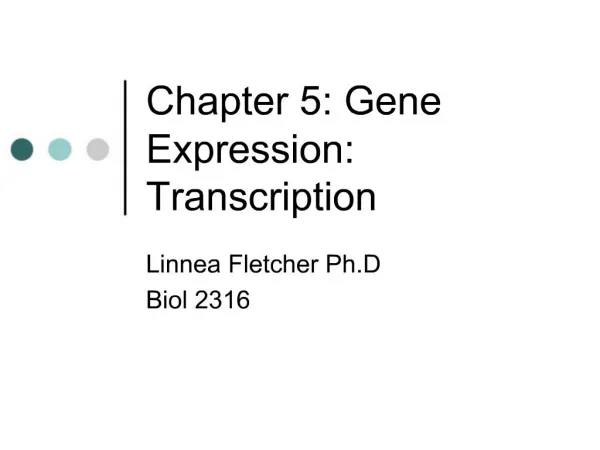
1. Chapter 5: Gene Expression: Transcription Linnea Fletcher Ph.D
Biol 2316
2. Yeast TBP (TATA binding protein), colored green, binding to a promoter region in DNA. Yeast TBP (TATA binding protein), colored green, binding to a promoter region in DNA.
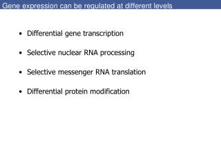
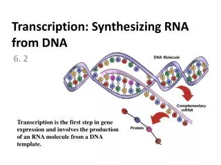
3. Principal Points DNA? RNA is transcription
Catalyzed by the enzyme RNA polymerase (does NOT require a primer)
There are similarities and differences in prokaryotic and eukaryotic transcription and the mRNA product
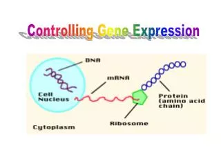
4. The Central Dogma DNA? RNA? Protein
Besides mRNA, What are the other types of RNA molecules that can be made?
rRNA
tRNA
snRNA includes iRNAs (involved in eukaryotic RNA processing)
(what do these RNAs do?)
5. Figure 5.1 Transcription process. The DNA double helix is denatured by RNA polymerase in prokaryotes and by other proteins in eukaryotes. RNA polymerase then catalyzes the synthesis of a single-stranded RNA chain, beginning at the �start of transcription� point. The RNA chain is made in the direction, with only one strand of the DNA used as a template to determine the base sequence. Figure 5.1 Transcription process. The DNA double helix is denatured by RNA polymerase in prokaryotes and by other proteins in eukaryotes. RNA polymerase then catalyzes the synthesis of a single-stranded RNA chain, beginning at the �start of transcription� point. The RNA chain is made in the direction, with only one strand of the DNA used as a template to determine the base sequence.
6. To Help You Understand the Differences and Similarities It might help you to make a table of the similarities and differences between DNA and RNA polymerases and between eukaryotes and prokaryotes
In prokaryotes, the RNA polymerase binds and opens the DNA while in eukaryotes, unwinding is done by other proteins that bind to the DNA at the start point for transcription
7. Figure 5.2 Chemical reaction involved in the RNA polymerase�catalyzed synthesis of RNA on a DNA template strand. Figure 5.2 Chemical reaction involved in the RNA polymerase�catalyzed synthesis of RNA on a DNA template strand.
8. Figure 5.3 Promoter, RNA-coding sequence, and terminator regions of a gene. The promoter is upstream of the coding sequence, the terminator downstream. Figure 5.3 Promoter, RNA-coding sequence, and terminator regions of a gene. The promoter is upstream of the coding sequence, the terminator downstream.
9. Figure 5.4 Action of E. coli RNA polymerase in the initiation and elongation stages of transcription. (a) In initiation, the RNA polymerase holoenzyme first binds loosely to the promoter at the -35 region. (b) As initiation continues, RNA polymerase binds more tightly to the promoter at the -10 region, accompanied by a local untwisting of about 17 bp around that region. At this point, the RNA polymerase is correctly oriented to begin transcription at +1(c) After eight to nine nucleotides have been polymerized, the sigma factor dissociates from the core enzyme. (d) As the RNA polymerase elongates the new RNA chain, the enzyme untwists the DNA ahead of it; as the double helix re-forms behind the enzyme, the RNA is displaced away from the DNA. Figure 5.4 Action of E. coli RNA polymerase in the initiation and elongation stages of transcription. (a) In initiation, the RNA polymerase holoenzyme first binds loosely to the promoter at the -35 region. (b) As initiation continues, RNA polymerase binds more tightly to the promoter at the -10 region, accompanied by a local untwisting of about 17 bp around that region. At this point, the RNA polymerase is correctly oriented to begin transcription at +1(c) After eight to nine nucleotides have been polymerized, the sigma factor dissociates from the core enzyme. (d) As the RNA polymerase elongates the new RNA chain, the enzyme untwists the DNA ahead of it; as the double helix re-forms behind the enzyme, the RNA is displaced away from the DNA.
10. Figure 5.5 Sequence of a Rho-independent terminator and structure of the terminated RNA. The mutations in the stem (yellow section) partially or completely prevent termination. Figure 5.5 Sequence of a Rho-independent terminator and structure of the terminated RNA. The mutations in the stem (yellow section) partially or completely prevent termination.
11. Transcription in Eukaryotes RNA pol I, located exclusively in the nucleolus, catalyzes the synthesis of 3 of the RNAs found in ribosomes: the 28S, 18S, and 5.8S
RNA pol II, found only in the nucleoplasm, synthesizes mRNAs and some snRNAs
RNA pol III, found only in the nucleoplasm, synthesizes the tRNAs, 5S rRNA, and snRNAs not made by pol II
12. Promoters and Enhancers How promoters are studied
Through mutational analysis
Compare DNA sequences upstream of a number of protein-coding genes
Found the following:
Promoters encompass about 200 bp upstream and contain two regions: the core promoter and promoter proximal elements
13. Core promoter: set of cis-acting sequence elements
Inr (Initiator) which spans the initiation start site (+1)
TATA box or TATA element located at -30 = TATAAA
Proximal Elements are further upstream (50 to 200 nucleotides); examples are as follows:
�cat� box (CAAT) -75; GC box -90
Enhancers (function upstream or downstream-1000 bp away)
The binding of Activator proteins to sequence elements
14. Figure 5.7 Assembly of the transcription initiation machinery. First, TFIID binds to the TATA box to form the initial committed complex. The multisubunit TFIID has one subunit called the TATA-binding protein (TBP), which recognizes the TATA box sequence, and a number of other proteins called TBP-associated factors (TAFs). In vitro, the TFIID�TATA box complex acts as a binding site for the sequential addition of other transcription factors. Initially, TFIIA and then TFIIB bind, followed by RNA polymerase II and TFIIF, to produce the minimal transcription initiation complex. (RNA polymerase II, like all eukaryotic RNA polymerases, cannot directly recognize and bind to promoter elements.) Next, TFIIE and TFIIH bind to produce the complete transcription initiation complex, also called the preinitiation complex (PIC). TFIIH�s helicase-like activity now unwinds the promoter DNA, and transcription is ready to begin. Figure 5.7 Assembly of the transcription initiation machinery. First, TFIID binds to the TATA box to form the initial committed complex. The multisubunit TFIID has one subunit called the TATA-binding protein (TBP), which recognizes the TATA box sequence, and a number of other proteins called TBP-associated factors (TAFs). In vitro, the TFIID�TATA box complex acts as a binding site for the sequential addition of other transcription factors. Initially, TFIIA and then TFIIB bind, followed by RNA polymerase II and TFIIF, to produce the minimal transcription initiation complex. (RNA polymerase II, like all eukaryotic RNA polymerases, cannot directly recognize and bind to promoter elements.) Next, TFIIE and TFIIH bind to produce the complete transcription initiation complex, also called the preinitiation complex (PIC). TFIIH�s helicase-like activity now unwinds the promoter DNA, and transcription is ready to begin.
15. Figure 5.8 shows the general structure of the mature, biologically active mRNA as it exists in both prokaryotic and eukaryotic cells. Figure 5.8 shows the general structure of the mature, biologically active mRNA as it exists in both prokaryotic and eukaryotic cells.
17. Figure 5.10 Cap structure at the end of a eukaryotic mRNA. The cap results from the addition of a guanine nucleotide and two methyl groups. Figure 5.10 Cap structure at the end of a eukaryotic mRNA. The cap results from the addition of a guanine nucleotide and two methyl groups.
18. Figure 5.11 Schematic diagram of the end formation of mRNA and the addition of the poly(A) tail to that end in mammals. In eukaryotes, the formation of the end of an mRNA is produced by cleavage of the lengthening RNA chain. CPSF binds to the AAUAAA signal, and CstF binds to a GU-rich or U-rich sequence (GU/U) downstream of the poly(A) site. CPSF and CstF also bind to each other, producing a loop in the RNA. CFI and CFII bind to the RNA and cleave it. Poly(A) polymerase then adds the poly(A) tail to which poly(A) binding proteins attach. Figure 5.11 Schematic diagram of the end formation of mRNA and the addition of the poly(A) tail to that end in mammals. In eukaryotes, the formation of the end of an mRNA is produced by cleavage of the lengthening RNA chain. CPSF binds to the AAUAAA signal, and CstF binds to a GU-rich or U-rich sequence (GU/U) downstream of the poly(A) site. CPSF and CstF also bind to each other, producing a loop in the RNA. CFI and CFII bind to the RNA and cleave it. Poly(A) polymerase then adds the poly(A) tail to which poly(A) binding proteins attach.
19. Figure 5.12 General sequence of steps in the formation of eukaryotic mRNA. Not all steps are necessary for all mRNAs. Most eukaryotic protein coding genes contain introns and some bacteriophage genes also contain introns.Figure 5.12 General sequence of steps in the formation of eukaryotic mRNA. Not all steps are necessary for all mRNAs. Most eukaryotic protein coding genes contain introns and some bacteriophage genes also contain introns.
20. Figure 5.13 Model for intron removal by the spliceosome. Spliceosomes consist of the premRNA bound to small nuclear ribonucleoprotein particles (snRNPs; also called snurps). SnRNPs are snRNAs associated with proteins and therre are 5 principal ones named U1, U2, U4, U5, and U6. Each one is associated with a number of proteins. At the end 5� of an intron is the sequence GU, and at the 3� end is the sequence AG. Near the end of the intron is an A nucleotide located within the branch-point sequence, which, in mammals, is YNCURAY, where Y=pyrimidine, N=any base, R=purine, A=adenine, and, in yeast, is UACUAAC. (The italic A in each sequence is where the 5� end of the intron bonds). With the aid of snRNPs, intron removal begins with a cleavage at the first exon�intron junction. The G at the released 5� of the intron folds back and forms an unusual 2�-5� bond with the A of the branch-point sequence. This reaction produces a lariat-shaped intermediate. Cleavage at the 3� intron�exon junction and ligation of the two exons completes the removal of the intron. Figure 5.13 Model for intron removal by the spliceosome. Spliceosomes consist of the premRNA bound to small nuclear ribonucleoprotein particles (snRNPs; also called snurps). SnRNPs are snRNAs associated with proteins and therre are 5 principal ones named U1, U2, U4, U5, and U6. Each one is associated with a number of proteins. At the end 5� of an intron is the sequence GU, and at the 3� end is the sequence AG. Near the end of the intron is an A nucleotide located within the branch-point sequence, which, in mammals, is YNCURAY, where Y=pyrimidine, N=any base, R=purine, A=adenine, and, in yeast, is UACUAAC. (The italic A in each sequence is where the 5� end of the intron bonds). With the aid of snRNPs, intron removal begins with a cleavage at the first exon�intron junction. The G at the released 5� of the intron folds back and forms an unusual 2�-5� bond with the A of the branch-point sequence. This reaction produces a lariat-shaped intermediate. Cleavage at the 3� intron�exon junction and ligation of the two exons completes the removal of the intron.
21. Figure 5.14 Self-splicing reaction for the group I intron in Tetrahymena pre-rRNA. Cleavage at the splice junction takes place first, accompanied by the addition of a G nucleotide to the end of the intron. Cleavage at the splice junction releases the intron; the two exons are spliced. The intron now circularizes by the nucleotide bonding to an internal nucleotide, producing a lariat-shaped molecule. Cleavage of the lariat molecule produces a circular RNA molecule and a linear molecule. Figure 5.14 Self-splicing reaction for the group I intron in Tetrahymena pre-rRNA. Cleavage at the splice junction takes place first, accompanied by the addition of a G nucleotide to the end of the intron. Cleavage at the splice junction releases the intron; the two exons are spliced. The intron now circularizes by the nucleotide bonding to an internal nucleotide, producing a lariat-shaped molecule. Cleavage of the lariat molecule produces a circular RNA molecule and a linear molecule.
22. Ribozymes RNAs that catalytically cleave RNA at specific sequences.
23. Figure 5.15 Comparison of the DNA sequences of the cytochrome oxidase subunit III gene (COIII) in the protozoans Trypanosome brucei (Tb), Crithridia fasiculata (Cf ), and Leishmania tarentolae (Lt), aligned with the conserved mRNA for Tb. The lowercase u�s are the U nucleotides added to the transcript by RNA editing. The template T�s in Tb DNA that are not in the RNA transcript are yellow. Figure 5.15 Comparison of the DNA sequences of the cytochrome oxidase subunit III gene (COIII) in the protozoans Trypanosome brucei (Tb), Crithridia fasiculata (Cf ), and Leishmania tarentolae (Lt), aligned with the conserved mRNA for Tb. The lowercase u�s are the U nucleotides added to the transcript by RNA editing. The template T�s in Tb DNA that are not in the RNA transcript are yellow.
24. Figure 5.16 Model of the complete (70S) ribosome of E. coli. The small (30S) ribosomal subunit is yellow, and the large (50S) ribosomal subunit is red. Figure 5.16 Model of the complete (70S) ribosome of E. coli. The small (30S) ribosomal subunit is yellow, and the large (50S) ribosomal subunit is red.
25. Figure 5.17 Composition of whole ribosomes and of ribosomal subunits in mammalian cells. Figure 5.17 Composition of whole ribosomes and of ribosomal subunits in mammalian cells.
26. Figure 5.18 rRNA genes and rRNA production in E. coli. (a) General organization of an E. coli rRNA transcription unit (an rrn region). (b) The scheme for the synthesis and processing of a precursor rRNA (p30S) to the mature 16S, 23S, and 5S rRNAs of E. coli. (The tRNAs have been omitted from this depiction of the processing scheme.) Figure 5.18 rRNA genes and rRNA production in E. coli. (a) General organization of an E. coli rRNA transcription unit (an rrn region). (b) The scheme for the synthesis and processing of a precursor rRNA (p30S) to the mature 16S, 23S, and 5S rRNAs of E. coli. (The tRNAs have been omitted from this depiction of the processing scheme.)
27. Figure 5.19 rRNA genes and rRNA production in eukaryotes. (a) Generalized diagram of a eukaryotic ribosomal DNA repeat unit. The coding sequences for 18S, 5.8S, and 28S rRNAs are indicated in green. NTS=nontranscribed spacer ETS=external transcribed spacer, ITS=internal transcribed spacer. (b) The transcription and processing of 45S pre-rRNA to produce the mature 18S, 5.8S, and 28S rRNAs. Figure 5.19 rRNA genes and rRNA production in eukaryotes. (a) Generalized diagram of a eukaryotic ribosomal DNA repeat unit. The coding sequences for 18S, 5.8S, and 28S rRNAs are indicated in green. NTS=nontranscribed spacer ETS=external transcribed spacer, ITS=internal transcribed spacer. (b) The transcription and processing of 45S pre-rRNA to produce the mature 18S, 5.8S, and 28S rRNAs.
28. Figure 5.20 Transfer RNA. Figure 5.20 Transfer RNA.