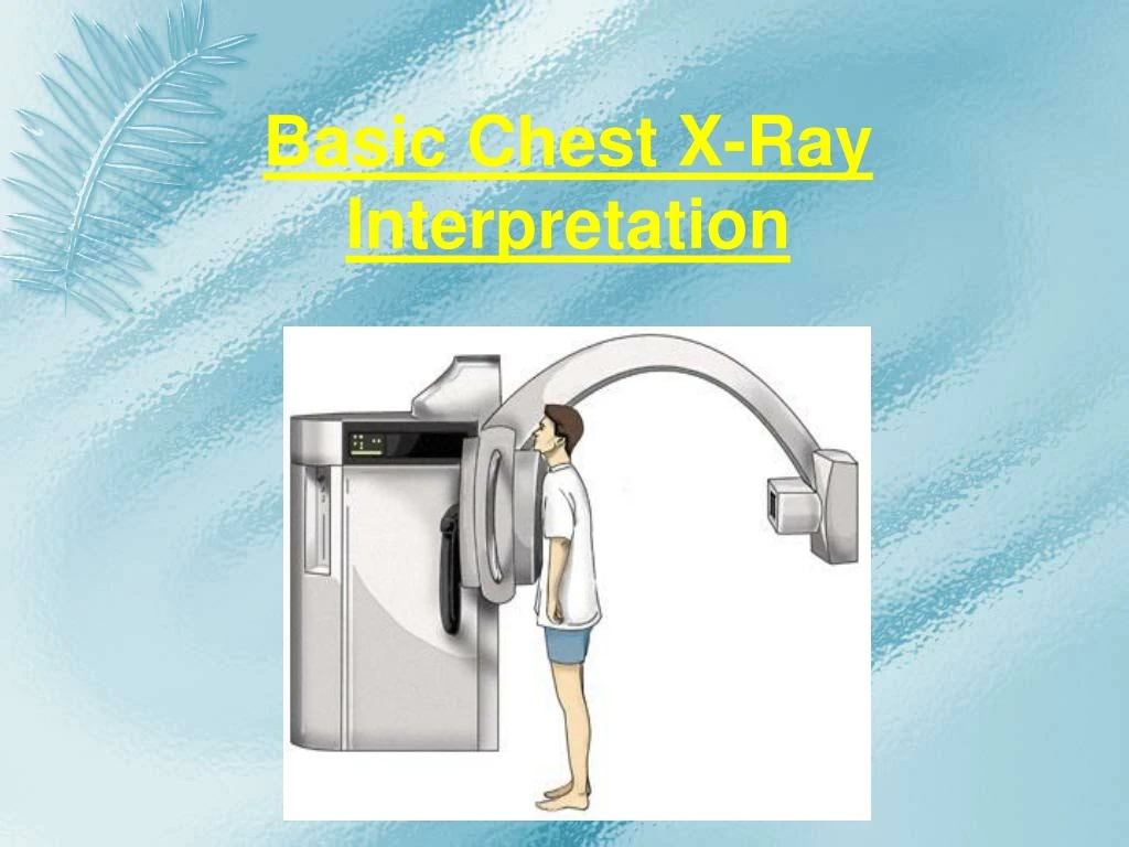

Comprehensive Guide to Basic Chest X-Ray and Arterial Blood Gas Interpretation
E N D
Presentation Transcript
Different tissues in our body absorb X-rays at different extents: • Bone- high absorption (white) • Tissue- somewhere in the middle absorption (grey) • Air- low absorption (black)
Systematic CXR Interpretation • IDENTIFICATION • TECHNIQUE • INTERPRETATION
Systematic CXR Interpretation • IDENTIFICATION • Correct patient • Correct date & time • Correct examination • Right vs. Left side (gastric bubble) • Comparison film.
Systematic CXR Interpretation • IDENTIFICATION • TECHNIQUE • INTERPRETATION
Systematic CXR Interpretation • TECHNIQUE • Complete exam • All views • Entire anatomical area included.
Systematic CXR Interpretation TECHNIQUE, cont. • Projection or Quality of the film: • First determine is the film a PA or AP view. • PA- the x-rays penetrate through the back of the patient on to the film. • AP-the x-rays penetrate through the front of the patient on to the film. The width of heart & mediastinum larger on AP film. • All x-rays in the ICU are portable and are AP view
Systematic CXR Interpretation TECHNIQUE, cont. • Position • Erect. • Supine. • Lateral position.
Systematic CXR Interpretation • TECHNIQUE, cont. • Penetration • Over-penetrated dark films can obscure subtle pathologies. • Under-penetrated white films may given impression of diffuse increased density.
Is the film over or under penetrated? • If under penetrated you will not be able to see the thoracic vertebrae.
Systematic CXR Interpretation • TECHNIQUE, cont. • Adequacy (full Inspiration) • Normal, erect, inspiratory CXR shows 9.5-10.5 posterior ribs. • Less inspiration appears diffusely denser • Diaphragms elevated causing heart & mediastinum to appear enlarged.
Systematic CXR Interpretation • TECHNIQUE, cont. • Rotation • Determine by observing the equal distance between the medial clavicular head and the spinous process of the thoracic vertebral body.
Systematic CXR Interpretation • IDENTIFICATION • TECHNIQUE • INTERPRETATION
Systematic CXR Interpretation • INTERPRETATION • Extraneous material • Contrast • Lines, tubes, clips • All properly located? • Bones • Fracture, dislocation • Mineralization • Soft tissues • Asymmetry • Calcifications
Systematic CXR Interpretation • INTERPRETATION • Diaphragms & Below • Free air • Dilated bowel • Abnormal position • Lung fields & mediastinum • Asymmetry , central mediastinum • Consolidation (opacity), nodule or lesion • Vascular marking. • Heart • Size & shape • Cardiothoracic ratio
Arterial Blood Gas Definition • Blood gases is a measurement of how much oxygen (O2) and carbon dioxide (CO2) is in your blood. • It also determines the acidity (pH) of your blood.
Arterial Blood Gas Why the Test is Performed? • To evaluate respiratory diseases and conditions that affect the lungs. • It helps determine the effectiveness of oxygen therapy.
Arterial Blood Gas How the Test is Performed? • Usually, blood is taken from an artery. • The blood may be collected from the radial artery in the wrist, the femoral artery in the groin, or the brachial artery in the arm. • May test circulation to the hand before taking a sample of blood from the wrist area. • Insert a small needle through the skin into the artery (You can use (anesthesia) applied to the site before the test begins).
Arterial Blood Gas How the Test is Performed • In rare cases, blood from a vein may be used. • After the blood is taken, pressure is applied to the site for a few minutes to stop the bleeding. • Watch the site for signs of bleeding or circulation problems. • The sample must be quickly sent to a laboratory for analysis to ensure accurate results.
Arterial Blood Gas How to Prepare for the Test • There is no special preparation. • If you are on oxygen therapy, the oxygen concentration must remain constant for 20 minutes before the test.
Arterial Blood Gas How the Test Will Feel • You may feel brief cramping or throbbing at the puncture site
Arterial Blood Gas Risks • There is very little risk when the procedure is done correctly. • Veins and arteries vary in size from one patient to another and from one side of the body to the other. • Taking blood from some people may be more difficult than from others.
Arterial Blood Gas Other risks associated with this test may include: • Bleeding at the puncture site • Blood flow problems at puncture site (rare) • Bruising at the puncture site • Delayed bleeding at the puncture site • Fainting or feeling light-headed • Hematoma (blood accumulating under the skin) • Infection (a slight risk any time the skin is broken)