Chapter 5 Electromagnetic Radiation
Chapter 5 Electromagnetic Radiation. A photon is the smallest element of electromagnetic energy. Photons are energy disturbances moving through space at the speed of light. Photons have no mass but they do have electric and magnetic fields. Electromagnetic Radiation.

Chapter 5 Electromagnetic Radiation
E N D
Presentation Transcript
Chapter 5 Electromagnetic Radiation • A photon is the smallest element of electromagnetic energy. • Photons are energy disturbances moving through space at the speed of light. • Photons have no mass but they do have electric and magnetic fields.
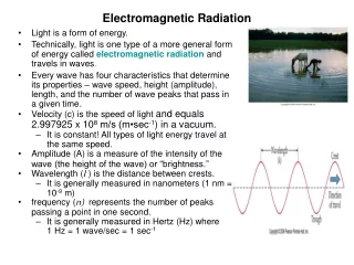
Electromagnetic Radiation • A field is an interaction between different energies, forces or masses that can not be seen but can be described mathematically. • Electromagnetic Radiation can be represented by the sine-wave model. • Sine-waves have amplitude.Amplitude is one half the range from crest to valley over a sine wave.
Electromagnetic Radiation • The important properties of the sine-wave model are frequency(f) and wavelength(λ) and velocity. • Frequency is the number of wavelengths passing a point per second. • Frequency is identified as oscillations per second and measured in hertz (Hz).
Electromagnetic Radiation • Wavelength is the distance from one crest to another or from any point in the wave to the next corresponding point. • The wave parameters are very important. A change in one affects the value of one or both of the others.
Electromagnetic Radiation • At a given velocity, wavelength and frequency are inversely proportional. • The Wave Formula • Velocity= Frequency x Wavelength
Electromagnetic Radiation • With EMF we know the velocity so the formula is simplified. • c= fλ or f= c/λ or λ= c/f • As frequency increases, wavelength decreases and vice versa • For electromagnetic radiation, frequency and wavelength are inversely proportional.
Electromagnetic Spectrum • The electromagnetic spectrum includes the entire range of electromagnetic radiation. • The frequency range is from about 102 to 1024 Hz • Photon wavelengths range from 107 to 10-16m. • Grouped together, these radiations make up the electromagnetic spectrum.
Electromagnetic Spectrum • Three important ranges. • Visible light • Radio frequency • X-radiation • Others include: • UV • IR and microwave
Electromagnetic Spectrum • EMF can be measured in three formats • Energy (eV) used to describe x-rays • Frequency (Hz) • Wavelength (m)
Visible Light • Measured in wavelength. • A prism is used to refract or change the direction of the photons. • Only form of EMF that we can sense.
Forms of Light • Visible light ranges from 700nm to 400nm wavelength. • Infrared light have longer wavelength than visible light but shorter than microwaves. • Ultraviolet light is located between visible light and ionizing radiation.
Radiofrequency • AM radio, FM radio and Television are other forms of electromagnetic radiation. • With radio, the frequency is used to identify the station. • Short wavelength radiofrequency are referred to as microwaves.
Ionizing Radiation • Unlike visible light or radiofrequency, ionizing electromagnetic radiation is characterized by the energy contained in the photon. • When we use 70 kVp, the photon will have energy varying from 0 to 70 keV.
Ionizing Radiation • The frequency is much higher and wavelength much shorter for x-rays compared to any other form of electromagnetic radiation. • Visible light identified by wavelength • Radiofrequency identified by frequency • X-rays identified by energy
Ionizing Radiation • The only difference between X-rays and gamma rays is their origin. • X-rays are produced outside the nucleus. • Gamma rays are produced inside the nucleus of radioactive atoms.
Wave-Particle Duality • A x-radiation photon and a visible light photon are fundamentally the same except that x-radiation photons have a much higher frequency and shorter wavelength. • These differences change the way they interact with matter. • Visible light tends to behave as waves.
Wave-Particle Duality • X-radiation tends to behave more as particles than waves. • Both types of photons exhibit both types of behavior and this is referred to as the wave-particle duality of radiation. • Photons interact with matter when the matter is approximately the same size as the photon wavelength.
Wave-Particle Duality • Radio & television photons wavelength is measured in meters and interact with long metal rods called antennae. • Microwave are measured in centimeters and react most easily with popcorn & hotdogs.
Wave-Particle Duality • Visible light wavelength is measured in micrometers or nanometers, interacts with living cells such as the rods and cones in the eye. • Ultraviolet light interacts with molecules. • X-rays interact with atoms and electrons. • All radiation with wavelengths longer than x-rays interact primarily as a wave.
Wave model: Visible Light • Vision is result of specially developed organ that sense a very narrow portion of the electromagnetic spectrum. • When a visible light photon strikes an object, it sets the molecule of the object into vibration.
Wave model: Visible Light • The orbital electrons become excited by the higher energy. This energy is immediately irradiated as another photon of light. This is referred to as reflection. • Atomic and molecular structure determine which wavelength of light are reflected.
Wave model: Visible Light • Light photons not reflected are either absorbed or transmitted. • There are three degrees of absorption: • Transparency • Translucency • Opacity
Degrees of Absorption • If all of the light is transmitted almost unaltered, it is transparent. • If only some of the light passes through , it is called translucent.
Degrees of Absorption • If all of the light is absorbed, it is called opaque. • Attenuation is the sum of scattering and and absorption of radiation.
Radiopaque or Radiolucent • Terms used to describe the appearance of objects on the x-ray film. • Objects that absorb the radiation are called radiopaque.
Radiopaque or Radiolucent • Structures that attenuate the x-rays are referred to as Radiolucent. • Bone is radiopaque. • Lung is radiolucent.
Inverse Square Law I1 D22 • ____ = _____ • I2 D12 • Radiation intensity is inversely related to the square of the distance from the source. • The decrease is due to the light being spread over a ever increasing area.
Inverse Square Law • If the source is not a point but a line such as a fluorescent lamp, the inverse square law does not hold at distances close to the source. • The inverse square law can be applied to distances greater than seven times the longest dimension of the source .
Inverse Square Law • The Inverse Square Law is used in radiography to adjust technical factors for changes in distance. • It is also used for radiation protection. The farther you are away from the source, the lower the exposure. • To use the formula, you need to know three of the 4 factors which are two distances and two intensities.
Particle Model: Quantum Theory • Unlike other forms of electromagnetic radiation, x-ray energy is measured in electron volts (eV). • X-ray energies range from 1 to 50 MeV • X-ray wavelengths range from 10-9 to 10-12m. • X-ray frequency range from 1018 to 1021Hz
Range of X-ray Energies • Diffraction uses less than 10 kVp. • Grenz rays with energies of 10 to 20 kVp are used in dermatology. • Diagnostic Radiography uses the range of 30 kVp to 150 kVp. • Therapy uses energies from 200 to 1000 kVp
X-ray Waveform • X-rays have both electric and magnetic fields. • One wave represents the electric field and one the magnetic field varying at right angles to each other.
Planck’s Quantum Theory • X-rays are created at the speed of light or they don’t exist at all. • The energy of a photon is directly proportional to it’s frequency. • A photon’s energy is inversely proportional to the photon wavelength. • E= hf where E is the photon energy; h is Planck’s constant or 10-15eVs or 6.63 x 10⁻³⁴Js and f is the photon frequency in hertz.
Matter and Energy • Like the law of the conservation of matter, the law of conservation of energy states that Energy can be neither created or destroyed. • Planck’s quantum physics and Einstein’s physics of relativity greatly extended these theories.
Matter and Energy • According to quantum physics and physics of relativity, matter can be transformed into energy and vise versa. • Although matter and energy are interchangeable, it is energy from the x-ray photon interacting with tissue and the image receptor that forms the basis of x-ray imaging.
Mass Energy Relationship • Mass and energy are two forms of the same medium. This scale shows the equivalence of mass measured in kilograms to energy measured in electron volts.
Summary • X-rays are just one type of photon of electromagnetic radiation • The following are used to describe the electromagnetic spectrum. • Frequency • Wavelength • Velocity • Amplitude • These factors are what determines such radiation interacts with matter.
X-Ray and Chiropractic • First used by B.J. Palmer at PSC in 1910. • He referred x-rays of the spine as spinographs. • Originally take to prove existence of the subluxations later used to evaluate spine for misalignment and pathology..
X-Ray and Chiropractic • 1924 first erect spinal radiographs at Universal Chiropractic College in Pittsburgh, Pa. This was the first time that the effects of gravity on the spine was evaluated. • Until 1938 all films taken at P.S.C. by Doctor Ray Richardson.
X-Ray and Chiropractic • Dr Warren Sausser was a 1917 graduate of Palmer School of Chiropractic. • He was a radiologic technologist in the Army during WW1. • He is one of the pioneers of radiography. Thanks to him Chiropractors were allowed to take x-rays.
X-Ray and Chiropractic • He is responsible for D.C.’s in New York being able to take x-rays. • He founded what is now the Chiropractic College of Radiology.
X-Ray and Chiropractic • He was the first chiropractor to take full body radiographs. • He also did extensive research in full spine radiography.
X-Ray and Chiropractic • Dr Hugh Logan stressed the importance of upright full spine films making the procedure popular. Dr. Sausser was impressed by his approach. • Dr Warren Sausser reported the first single exposure full spine taken with the 14” x 36” film. Standard X-ray Company designed the x-ray machine . Kodak developed cassette, film and the special hangers needed to process the film. The cassette weighed 50 pounds.
X-Ray and Chiropractic • Dr. Sausser developed filters to equalize the exposure. • The tube was placed nine feet from the patient and a 12 second exposure was needed to produce the image. • Dr. Sausser also developed full body exams but only the full spine remains today. • The full body radiograph went the way of the x-rays to fit shoes.
X-Ray and Chiropractic • Like many of the early pioneers of x-ray, he died in 1958 as a result of the affects of radiation exposure. • He had tumors down his side that was not behind a barrier during the exposures. • He would often peek around the barrier to check on the patient.
X-Ray and Chiropractic • Films taken recumbent until 1938 at Palmer School of Chiropractic. • All forms of radiography was done at P.S.C. including fluoroscopic contrast studies of G.I. Tract and gallbladder until 1938.
X-Ray and Chiropractic • The use of radiography started as a means to see the subluxations. Today it is used to image the entire body and not just the spine. • Today the scope is generally limited to plain film radiography of the spine, chest, abdomen and extremities.
X-Ray and Chiropractic • As early as 1922, they used x-rays for the detection of pathological processes, fractures and anomalies that impact the patients health as well as chiropractors determinations of what to do or not to do. • This is still true today.
End of Lecture Return to Physics Lecture Index Return to LC-232 Principles & Physics Homepage