Pregnancy Diagnosis Prenatal Diagnosis
Pregnancy Diagnosis Prenatal Diagnosis. Obstetrics and Gynecology Hospital of Fudan University Xing Chen, MD. Email: xing_chen@21cn.com. Pregnancy Diagnosis.

Pregnancy Diagnosis Prenatal Diagnosis
E N D
Presentation Transcript
Pregnancy DiagnosisPrenatal Diagnosis Obstetrics and Gynecology Hospital of Fudan University Xing Chen, MD. Email: xing_chen@21cn.com
For a woman with regular menstrual cycles, a history of one or more missed periods following a time of sexual activity without effective contraception strongly suggests early pregnancy
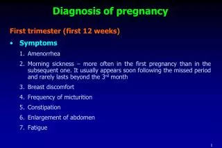
Signs and symptoms of early pregnancy • Amenorrhea • Nausea with or without vomiting • Breast enlargement and tenderness • Increased frequency of urination without dysuria • Fatigue
Physical Examination • softening and enlargement of the pregnant uterus • congestion and a bluish discoloration of the vagina (Chadwick sign) • softening of the cervix (Hegar sign) • Increased pigmentation of the skin • appearance of circumlinear striae on the abdominal wall (striae gravidarum) • palpation of fetal parts • appreciation of fetal movement and fetal heart tones
human chorionic gonadotropin (hCG): α/β-subunit produced in the syncytiotrophoblast Urine approximately 4 weeks following the first day of the last menstrual period All urine pregnancy tests are best performed on early-morning urine specimens, which contain the highest concentration of hCG Serum specific and sensitive by following serial quantitative hCG levels and comparing them to the expected rise derived from normative data for proven normal intrauterine pregnancies Pregnancy test
In most normal pregnancies at hCG levels below 1,200 mIU/ml the hCG usually doubles every 48-72 HOURS and it normally increases by at least 60% every TWO DAYS
Ultrasound examination • Abdominal ultrasound allowing visualization of a normal pregnancy gestational sac 5 to 6 weeks after the beginning of the last normal menstrual period (corresponding to β-hCG concentrations of 5000 to 6000 mIU/mL) • Transvaginal ultrasoundoften detects pregnancy at 3 to 4 weeks of gestation (corresponding to β-hCG concentrations of 1000 to 2000 mIU/mL)
Image of an early gestational sac containing a yolk sac and early embryo. The yolk sac is the circular hyperechoic structure adjacent to the embryo. Image of an early gestational sac demonstrating the early embryo. Calipers are placed at both ends of the embryo measuring the longest length from the "crown to the rump" giving the crown-rump length. This measurement is used for dating the pregnancy.
Acoustic fetoscope beyond 18 to 20 weeks of gestational age Electronic Doppler devices approximately 12 weeks of gestation Detection of fetal heart activity “fetal heart tones”
Diagnosis • Detection of human chorionic gonadotropin (hCG) in blood or urine • Identification of pregnancy by ultrasound examination • Identification of fetal cardiac activity by Doppler ultrasound
Abnormal Pregnancy • Spontaneous abortion • Ectopic pregnancy • Trophoblastic disease
SUMMARY • The most common signs and symptoms of pregnancy are amenorrhea, nausea/vomiting, breast tenderness, urinary frequency, and fatigue • The most sensitive method of detecting hCG is a serum pregnancy test • Almost all pregnant women will have a positive urine pregnancy test one week after the first day of a missed menstrual period
SUMMARY • The diagnosis of early pregnancy is based primarily upon laboratory assessment of human chorionic gonadotropin (hCG) • Identification of pregnancy by ultrasound examination or identification of fetal cardiac activity by Doppler ultrasound are alternative methods, but sensitivity depends on the gestational age • Transvaginal ultrasound examination can visualize a gestational sac at 4.5 to 5 weeks of gestation
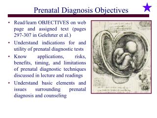
Detect genetic disorders and birth defects • > 200 single gene disorders can be diagnosed • Testing done only when a family history or other risk
Prenatal diagnosisis the science of identifying structural or functional abnormalities-birth defects-in the fetus
Etiology of Birth Defects • Malformation • Deformation • Disruption • Other
Malformation • an intrinsic abnormality "programmed" in development, regardless of whether a precise genetic etiology is known • spina bifida
Deformation • caused when a genetically normal fetus develops abnormally because of mechanical forces imposed by the uterine environment • normal limb that develops contractures because of prolonged oligohydramnios
Disruption • which is a more severe change in form or function that occurs when genetically normal tissue is modified as the result of a specific insult • an amnionic band causing a cephalocele or limb-reduction abnormality
Other • Syndrome: trisomy 18 • Sequence: oligohydramnios leading to pulmonary hypoplasia • Association: VATER vertebral defects anal atresia tracheoesophageal fistula with esophageal atresia radial dysplasia
Techniques • Non-invasive • Minimally invasive • Invasive
Non-invasive techniques • Ultrasound • Magnetic Resonance Imaging (MRI)
Ultrasound Noninvasive, uses reflected sound waves converted to an image Transducer placed on abdomen See physical features of fetus, not chromosomes May ID some chromosomal abnormalities by physical features
Minimally Invasive Techniques • Cell free fetal DNA (cffDNA) • Pre-implantation genetic diagnosis (PGD)
Fetal Cells in Maternal Circulation • Types • Placental cells • White blood cells • Immature red blood cells with nuclei • Enter the bloodstream (~6 and 12 weeks) • Fetal cells, only 1/100,000 in mother’s blood • Techniques need to be developed
Preimplantation Genetic Diagnosis (PGD) Eggs collected, fertilized, allowed to develop ~Third day of fertilization, embryo has 6–8 cells For PGD, one cell, a blastomere, is removed DNA extracted and tested Embryo without genetic disorder are implanted into mother
Invasive Techniques • Chorionic villus sampling (CVS) • Amniocentesis • Percutaneous umbilical blood sampling (cordocentesis)
Amniocentesis • Diagnose > 100 disorders, cells analyzed for chromosomal and biochemical disorders • Risk of infection and spontaneous abortion • Normally only used when: • Advanced maternal age • History of chromosomal disorder • Parent with chromosomal abnormality • Mother carrier of X-linked disorder
Removal of about 20 ml of amniotic fluid containing suspended cells that were sloughed off from the fetus Biochemical analysis of the amniotic fluid after the fetal cells are separated out Centrifugation Fetal cells are removed from the solution Analysis of fetal cells to determine sex Cells are grown in an incubator Karyotype analysis p. 46
Chorionic Villus Sampling (CVS) • Done for similar reasons as amniocentesis • Performed earlier than amniocentesis • 6–10 weeks vs. 16 weeks • Karyotypes available within a few hours or days • Increased risk of spontaneous abortion (0.5–2%)
Neural-Tube Defects (NTDs) • anencephaly, spina bifida, cephalocele, and other rare spinal fusion (schisis) abnormalities • had higher levels of alpha-fetoprotein (AFP) in maternal serum and amnionic fluid
Maternal Serum AFP Screening influence factors: maternal weight, gestational age, diabetes, multifetal gestation
Alpha-fetoprotein (AFP) • Glycoprotein of unknown function • Used to screen for open NTDs • 15-22 weeks gestation • Detection rate 80-85% • Used to screen for trisomy 21 • 15-20 weeks gestation • Detection rate 20-25%
Evaluation of Maternal Serum AFP Elevation • genetic counseling • diagnostic test • Specialized Sonography • amniocentesis
Specialized Sonography • Transverse and sagittal images of the spine are increasingly used to characterize the size and location of spinal defects
anencephaly spina bifida cephalocele