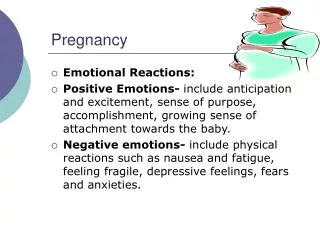
PREGNANCY
PREGNANCY. Prof. Aziza Tosson. Outline. Fertilization Development before Birth Fetal Circulation Embryonic Development Fetal Development Birth. Conception. A secondary oocyte can be fertilized for about 24 hours after ovulation

PREGNANCY
E N D
Presentation Transcript
PREGNANCY Prof. Aziza Tosson
Outline • Fertilization • Development before Birth • Fetal Circulation • Embryonic Development • Fetal Development • Birth
Conception • A secondary oocyte can be fertilized for about 24 hours after ovulation • Sperm remain viable for up to 48 hours within the female reproductive tract • This gives a three day “window” for intercourse to result in fertilization: two days before to one day after ovulation
Fertilization • Steps of fertilization (Conception). • Several sperm penetrate corona radiata. • Several sperm attempt to penetrate zona pellucida. • One sperm enters egg and nuclei fuse, producing a zygote. • Egg’s plasma membrane and zona pellucida change to prevent polyspermy.
Fertilization usually takes place in the outer one-third of the uterine tube, but can take place in the abdominal cavity • Sperm swim up the female reproductive tract, aided by muscular contractions of the uterus stimulated by prostaglandins in the semen. • The oocyte may also secrete a chemical that attracts sperm
Occurrence of Pregnancy • When a zygote begins dividing, it is termed an embryo. • Developing embryo travels down oviduct and eventually implants in endometrium. (Implantation / pregnancy) • Presence of human gonadotropic hormone (HCG) in the blood confirms pregnancy. • If implantation does not occur, a woman never knows fertilization took place.
Zygote undergoes rapid mitotic cell division, but these do not increase the size of the zygote – called cleavage divisions • Cleavage produces a solid sphere of cells, still surrounded by zona pellucida – now called a morula. • At 4.5 to 5 days, cells have developed into a hollow ball of cells – blastocyst. • It is at this stage that it enters the uterus.
Cellular differentiation Embryonic membranes Amnion – inner layer Chorion – outer layer Both membranes form bag of waters Amniotic fluid 700-1000 cc normal < 400 ml oligohydramnios > 2000 ml polyhydramnios Fetus contributes to amniotic fluid volume thru fetal urine Amniotic fluid important for fetal lung development Wajed Hatamleh RN, MSN, PhD.
Cellular differentiation Umbilical cord Wharton’s jelly 2 arteries/ 1vein Placenta Site of metabolic and nutritional exchange Maternal side – decidua basalis Fetal side – amnion 15 – 20 cotyledons: contain the complex vascular system of villi Wajed Hatamleh RN, MSN, PhD.
Implantation • The blastocyst remains free in the uterus a short time, during which the zona pellucida disintegrates. • Blastocyst nourished by glycogen from glands of the endometrium. • At about 6 days after ovulation blastocyst implants – orients cell mass toward endometrium, and secretes enzymes which allow it to penetrate (digest) the endometrial wall. This nourishes the blastocyst for about a week after implantation.
Following implantation, the placenta • originates from maternal and fetal • tissues. • Placenta then produces human chorionic • gonadotropin (HCG) which maintains the • corpus luteum in the ovary until the • placenta begins its own production of • progesterone and estrogen. • Physical signs of pregnancy include : no • menstruation, increased urination, • morning sickness, increased size of • breasts, and darkening of areolae.
As early as 8 -12 days after fertilization, the blastocyst begins to secrete human chorionic gonadotropin or hCG. • hCG keeps the corpus luteum active until the placenta can produce estrogens and progesterone. • The presence of hCG is the basis for pregnancy tests.
Inner cell mass forms two cavities: • The yolk sac • Amniotic cavity • In humans the yolk sac produces blood cells and future sex cells • The amniotic cavity becomes the cavity in which the embryo floats. Fluid is produced from fetal urine, and secretions from the skin, respiratory tract, and amniotic membranes.
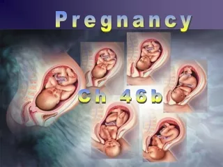
Gestation period • Divided into three trimesters. • During first trimester individual starts out as a zygote, then morula, blastocyst, and after implantation, is called an embryo. • Embryonic phase of development lasts from fertilization until the 8th week of gestation, when it becomes a fetus. • By day 35 the heart is beating, and eye and limb buds are present.
Fetus at 14 and 20 weeks gestation Wajed Hatamleh RN, MSN, PhD.
By month four, the rudiments of all organ systems are formed and functioning, and from then on, fetal development is primarily a matter of growth. • By the end of the third month the placenta is functioning.
Fifth through Seventh Months • Mother begins to feel fetal movement. • Wrinkled skin covered by fine hair, lanugo, is covered by a greasy substance vernix caseosa. • Lungs lack surfactant so if baby is born prematurely it will have to be on a respirator (respiratory distress syndrome).
The placenta • The chorion develops into the fetal part of the placenta. • The chorionic villi connect the fetal circulation to the placenta • Composed of both fetal and maternal tissues
Functions of the placenta: 1 Transfer gasses 2 Transport nutrients 3 Excretion of wastes 4 Hormone production – temporary endocrine organ – estrogen and progesterone 5 Formation of a barrier – incomplete, nonselective – alcohol, steroids, narcotics, anesthetics, some antibiotics and some organisms can cross
Placental functions Simple diffusion - water, oxygen, carbon dioxide, sodium and chloride Facilitated transport – glucose and galactose Active transport – amino acids, calcium, iron, iodine, vitamins, and glucose Pinocytosis – albumin and gamma globulin Hydrostatic and osmotic pressure Endocrine – hCG, hPL, estrogen and progesterone Wajed Hatamleh RN, MSN, PhD.
HORMONES OESTROGEN • Produced in corpus luteum • Produced by placenta after 12 weeks • Responsible for growth particularly of uterus and breasts
progesterone • Produced in corpus luteum and then the placenta • Relaxes smooth muscle • Inhibits uterine contractions until uterus is prepared for labour • Regulates storage of body fat
Human chorionic gonadotrophic • Secreted from trophoblast of the developing embryo • Maintains corpus luteum until placenta takes over • Used in tests to confirm pregnancy
Human placental lactogen • Alters maternal metabolism • Diverts glucose to fetus • Mobilises free fatty acids from maternal stores
RELAXIN • Released by corpus luteum then the Placenta • Softens pelvic ligaments • Reduces myometrial tone