Download
1 / 20
290 likes | 828 Vues
Medically Important Gram negative bacilli (Part 2). Dr Ekta Chourasia Lecturer, Microbiology. 1. Gram-negative bacilli a. Family Enterobacteriaceae: Medically important species Escherichia coli , S almonella typhi , Shigella, Klebsiella sp, Proteus sp Yersinia sp

E N D
Medically Important Gram negative bacilli (Part 2) Dr Ekta Chourasia Lecturer, Microbiology
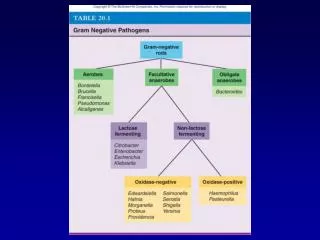
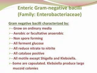
1. Gram-negative bacilli a. Family Enterobacteriaceae:Medically important species Escherichia coli, Salmonella typhi, Shigella, Klebsiella sp, Proteus sp Yersinia sp b. Pseudomonas sp, relatedOrganisms c. Bordetella pertussis d. Haemophilus spp 2. Curved bacilli Vibrio spp Campylobacter spp Helicobacter spp 3. Miscellaneous Legionella spp Chlamydia spp Rickettsia Representative Gram-negative Bacilli Dr Ekta Page 2
Vibrio cholerae • Gram-ve slender bacilli, comma shaped. • Actively motile by a long polar flagellum - Darting motility. • The causative agent for cholera. • Transmission – food & water contaminated with feces of patients & carriers. Dr Ekta Page 3
Pathogenicity • Cholera – acute diarrhoeal disease, passage of large volumes of rice watery stools, dehydration, hypovolemic shock & metabolic acidosis. • ID – 1011 org/ ml • Cholera toxin (CT) – endotoxin (same as E.coli LT) • Toxin coregulated pilus (TCP) Dr Ekta Page 4
Cholera Toxin (CT) • Resembles heat labile toxin of E.coli in its structure, function and mode of action • Complex of polypeptide subunits. • LT: one subunit of A (action- enzymic), five subunits of B (binding) Dr Ekta Page 5
Cholera Toxin (CT) Escherichia coli / Vibrio cholerae Gut lumen Intestinal epithelial cell Dr Ekta Page 6
Laboratory Diagnosis • Specimen – Feces, mucus flakes– before giving antibiotics. • Microscopy • Motility: darting motility • Gram stain: Gram –ve curved bacilli • Culture – Aerobic, alkaline pH (> 8). • Transport media – Cary Blair media, VR (Venkataraman-Ramakrishnan) media • Enrichment media – Alkaline Peptone Water, Monsur’s taurocholate tellurite PW. • Selective media – Alk. bile salt agar (BSA), TCBS (thiosulfate, citrate, bile salts & sucrose) agar. TCBS Agar Dr Ekta Page 7
Laboratory Diagnosis • Biochemical reactions • Oxidase +ve • Ferments sucrose, late lactose fermenter • ‘String test’ +ve ( with 0.5% sodium deoxycholate) • ‘Cholera red’ reaction – conc. Sulphuric acid to peptone water culture. • Slide agglutination – initially with cholera O subgroup I serum, then test for specific serotypes. • Immobilisation test – using antiserum, rapid test • Phage typing – NICED, Calcutta (INDIA). Dr Ekta Page 8
Prophylaxis & Treatment • Oral vaccines – immunity lasts for 6- 12 months. • Oral Rehydration fluid • Antibiotic therapy of secondary importance. Dr Ekta Page 9
Campylobacter jejuni • Curved or spiral Gram negative bacilli • Micro-aerophilic, grows best at 42oC • Symptoms develop within 2-5 days after exposure: • diarrhoea, cramping, abdominal pain • malaise • fever and • can include nausea and vomiting. • Usually self-limiting • Sources - Raw or undercooked meat and poultry, contaminated water or milk. • Prevention - Proper cooking, milk pasteurising and chlorination of water Dr Ekta Page 10
Helicobacter pylori • Curved or spiral Gram negative rods • Inflammation of gastric mucosa – leads to peptic ulcer • Produces abundant “urease” enzyme • rapid diagnostic test in gastric biopsy samples • ‘Urease breath test’ Dr Ekta Page 11
Hemophilus – General features • Part of normal flora in the upper respiratory tract or vagina • Morphology – pleomorphic, small, Gram-ve coccobacilli. • Cultural characteristics - fastidious • Requires about 10% CO2 • Enriched media – BA (special techniques) , CA • Accessory growth factors – X & V, present in blood. X - hemin, heat stable V – NAD, heat labile Dr Ekta Page 12
Pathogenicity of Hemophilus sps Pink eye (infectious conjunctivitis) Dr Ekta Page 13
Hemophilus influenzae • Strains isolated from acute infections are mainly capsulated (polysaccharide) • Six capsular types – a to f • Type b is associated with most of acute invasive infections like meningitis. • Capsulated strains – mainly infects children from 2 months to 3 yrs old • Transmission – respiratory route • Hib vaccine- protects against H. influenzae type b infections. Dr Ekta Page 14
Laboratory diagnosis • Specimen – CSF, blood, sputum • Microscopy – gram –ve bacilli that do not stain well • Culture – • CSF should bedirectly plated on BA, CA • Blood culture – positive for laryngoepiglottitis, pneumonia • Selective media – bacitracin added to CA – inhibits many Gram+ve bacteria found in URT • Special tests – • Satellitism – streak of S. aureus across the plate (BA or CA) provides V factor & enlarges the size of the colony. • Growth in presence of X & V factor S. aureus V XV X Dr Ekta Page 15
Bordetella pertussis • Commensal of the human respiratory tract. • Morphology – • Short, sometimes oval, Gram-ve bacilli (coccobacilli). • Freshly isolated strains may be capsulated. • Causes pertussis or whooping cough, a communicable childhood disease • Reservoir: apparently healthy carriers • Transmission by direct contact or inhalation of aerosols • Pertussis toxin: destroy & dislodge ciliated cells, responsible for whooping cough, can be toxoided Dr Ekta Page 16
Pathogenesis & Pathogenicity • Pertussis / Whooping cough – in pre school children. • loss of ciliary mechanism leads to buildup of mucus & blockage of the airways • hacking coughs followed by abrupt deep inhalation (whoop) • Characterised by 3 stages • Catarrhal - maximum infectivity • Paroxysmal - continuous cough • Convalescent - frequency & severity of cough decreases • Complications • Subconjunctival h’ge • Bronchopneumonia, lung collapse. • Convulsions, coma • Prophylaxis & Treatment • Immunisation – DPT • Children below 4 yrs should receive booster & chemoprophylaxis (erythromycin) if they come in contact with cases • antibiotics beneficial only within first 10 days of disease. Dr Ekta Page 17
Laboratory Diagnosis • Specimen - Postnasal swab, Pernasal swab • Cough plate method • Microscopy: gram-ve coccobacilli • Culture: Aerobic, Grows well on • Charcoal BA (with Pn or Cephalexin). • Bordet- Gengou medium(potato-glycerol-blood agar): ‘split pearls’ or ‘mercury drops’ like colonies. • Serological diagnosis not of much use. Dr Ekta Page 18
Yersinia pestis • Nonenteric…tiny, gram-negative bacilli (bipolar staining - safety pin appearance) • Causes Plague – three types: • Bubonic: involves lymph nodes of groin & axillae, called “bubos” • Pneumonic: infection localized to lungs, highly contagious; fatal without treatment • Septicemic: intravascular coagulation & subcutaneous hemorrhage. • Transmitted by rat flea Dr Ekta Page 19
Legionella pneumophila • Thin, non-capsulated Gram-ve coccobacilli • widely distributed in water ( mostly air-conditioners, cooling systems) • Causes Legionnaires disease (atypical pneumonia) & Pontiac fever Dr Ekta Page 20

![GRAM NEGATIVE BACILLI- MICRO {ST1]](https://cdn1.slideserve.com/2240310/gram-negative-bacilli-micro-st1-dt.jpg)