Download

1 / 30
380 likes | 978 Vues
Medically Important Gram negative bacilli (Part 1). Dr Ekta Chourasia Lecturer, Microbiology. 1. Gram-negative rods a. Family Enterobacteriaceae: Medically important species Escherichia coli , S almonella typhi , Shigella sp, Klebsiella spp, Proteus spp Yersinia spp.

E N D
Medically Important Gram negative bacilli (Part 1) Dr Ekta Chourasia Lecturer, Microbiology
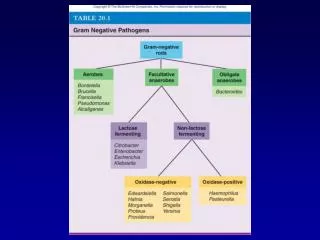
1. Gram-negative rods a. Family Enterobacteriaceae: Medically important species Escherichia coli, Salmonella typhi, Shigella sp, Klebsiella spp, Proteus spp Yersinia spp. b. Pseudomonas sp. & related organisms c. Bordetella pertussis d. Haemophilus spp 2. Curved rods Vibrio spp Campylobacter spp Helicobacter spp 3.Miscellaneous Legionella spp Chlamydia spp Rickettsia Representative Gram-negative Bacilli Dr Ekta Page 2
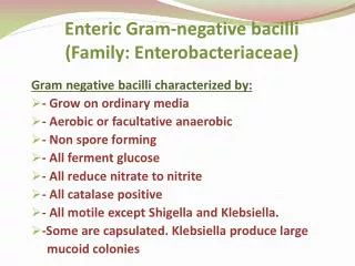
General Features of Enterobacteriaceae Present in large intestine Gram negative bacteria Aerobic or facultative anaerobic Motile by peritrichate flagella Grow on ordinary media (non fastidious) Ferments glucose with acid & gas or only acid Catalase + ve & oxidase -ve Dr Ekta Page 3
Classification of Enterobacteriaceae • Based on lactose fermentation – oldest method : • Lactose fermenters e.g. Escherichia coli, Klebsiella sps • Late lactose fermenters e.g. Shigella sonnei • Non lactose fermenters e.g Salmonella, Shigella sps other than Shigella sonnei • Commensal intestinal bacteria: LF • Intestinal pathogens: NLF NLF LF Dr Ekta Page 4
Escherichia coli • Named after Escherich, first to describe colon bacillus • Normal flora of the human & animal intestine. • Remains viable in the feces for few days. • Detection of E. coli in the drinking water – indicates recent pollution with human or animal feces. Dr Ekta Page 5
Antigenic Structure of Gram –ve Bacteria • Three antigens – serotyping of E.coli • H – flagellar antigen • O – somatic antigen • K – capsular antigen Majority do not possess K Ag. Dr Ekta Page 6
Virulence Factors - Two types of virulence factors: Surface Ags & Toxins • Surface Antigens • LPS surface O Ag – endotoxic activity • Envelope or K Ag – protects against phagocytosis • Fimbriae – colonisation, found in strains causing diarrhoea and urinary tract infections • Toxins (Exotoxins) – two types • Enterotoxins – pathogenesis of diarrhoea - 3 types : LT (heat labile toxin), ST (heat stable toxin) & VT (verocytotoxin or shiga- like toxin) • Hemolysins Dr Ekta Page 7
Heat Labile Toxin (LT) • Resembles cholera toxin in its structure, function and mode of action • Complex of polypeptide subunits. • LT: one subunit of A (action- enzymic), five subunits of B (binding) Dr Ekta Page 8
Heat Labile Toxin (LT) Escherichia coli / Vibrio cholerae Gut lumen Intestinal epithelial cell Dr Ekta Page 9
Pathogenicity / Clinical Infections • Urinary tract infection • Diarrhoea • Infantile diarrhoea • Traveller’s diarrhoea • Bloody/ Hemorrhagic diarrhoea • Pyogenic infections • Wound infection, especially after surgery of lower intestinal tract. • Peritonitis. • Biliary tract infection. • Neonatal meningitis. • Septicemia – can lead to fatal conditions like Septic shock Dr Ekta Page 10
Lab Diagnosis of UTI Specimens Urine Mid stream urine (MSU) Catheter specimen urine (CSU) Supra pubic aspiration (SPA) Microscopy Wet mount Pus cells / hpf Bacteria Gram stain Gram negative bacteria (1bacteria / oil field is significant) Urine Culture To know significant bacteriuria Dr Ekta Page 11
Lab Diagnosis of E. coli UTI Significant bacteriuria > 105 organism / ml of MSU Culture BA / MAC : LF (flat) CLED medium Identification tests I M Vi C test: + + - - Acid, no gas TSI agar Dr Ekta Page 12
Diarrheagenic E.coli • Enteropathogenic E. coli (EPEC) – infantile diarrhea, nontoxigenic • Enterotoxigenic E. coli (ETEC) – traveller’s diarrhea, resembles cholera • Enteroinvasive E. coli (EIEC) – bloody diarrhea (blood, mucus & leucocytes with stool) • Enterohemorrhagic E. coli (EHEC) or Verocytotoxigenic E. coli (VTEC):- O157:H7 serotype (food poisoning) - Hemorrhagic colitis, Hemolytic uraemic syndrome • Enteroaggregative E. coli (EAEC) : “stacked brick” appearance- persistent diarrhea in children • Diffusely adherent E. coli (DAEC) Dr Ekta Page 13
Klebsiella pneumoniae • General features • Normal gut flora in the intestine • Gram negative bacilli (short & plump) • Capsulated, non-motile, produces mucoid LF colonies on MAC • Pathogenicity • Pneumonia: hospital & community acquired • Meningitis & enteritis in infants • Urinary Tract Infection • Septicemia Dr Ekta Page 14
Proteus Normal gut flora in the intestine Gram negative bacilli, pleomorphic Motile, Non lactose fermenter (NLF) on MAC Swarms on BA, Urease +, H2S + Species P. mirabilis P. vulgaris UTI Pneumonia Wound infections Urease converts urea to NH4 & CO2 causing alkalinization of urine leading to renal calculi (stones) Proteus antigens are used in the Weil - Felix test to diagnose Rickettsial diseases Dr Ekta Page 15
Shigella • Classification – 4 species : biochemical & serological characteristics. - Sh.dysenteriae - Sh.flexneri Non Lactose Fermenter - Sh.boydii - Sh.sonnei - Late Lactose Fermenter Dr Ekta Page 16
Shigella species- Mannitol fermentation Mannitol Non Fermentation Fermentation S. dysenteriae - 12 S. flexneri- 6 S. boydii - 18 S. sonnei (Late lactose fermenter) Dr Ekta Page 17
Epidemiology & Clinical Syndromes • Causes Bacillary Dysentery – frequent passage of blood stained, mucopurulent stools. • Incubation period: 1-7 days, usually 48 hrs • Low Infectious dose: 10-100 bacilli • Feco-oral transmission • Common in pediatric age group (1-10 years) – leading cause of infantile diarrhea. • Sh.dysenteriae type I : most serious form of dysentery. • Shigellosis : whole spectrum of disease caused by Shigella. • Complication: Hemolytic Uremic Syndrome Dr Ekta Page 18
Pathogenesis Two-stage disease Early stage Watery diarrhea attributed to the enterotoxin activity ofShiga toxin inthe small intestine Second stage Dysenterydue toadherence and tissue invasion of large intestine(cytotoxic activity of Shiga toxin) Fever attributed toneurotoxic activity of toxin Shiga toxin Enterotoxic, neurotoxic and cytotoxic Similar to Shiga-like toxin of Enterohemorrhagic E. coli(EHEC) Dr Ekta Page 19
Laboratory Diagnosis • Specimen: fresh feces – mucus flakes (buffered glycerol saline – transport medium) • Microscopy: Gram–ve, nonmotile bacilli • Culture • MacConkey agar: NLF colonies • Enrichment broth – Selenite F, Gram-ve broth • Selective media – Deoxycholate agar (DCA), Salmonella-Shigella (SS) agar, XLD (Xylose Lysine deoxycholate) • Slide agglutination with polyvalent & monovalent sera. Treatment • Oral rehydration • Antibiotics for severe & toxic cases – Nalidixic acid or Norfloxacin. Dr Ekta Page 20
Salmonella • Gut of domestic animals & poultry. • Divided into 2 groups : • Enteric fever group – typhoid & paratyphoid bacilli. • Food poisoning group – usually animal parasites, producing gastroenteritis, septicemia or localized infections. Dr Ekta Page 21
Pathogenesis • Source of infection - Carriers , Cases, Poultry, dairy • Transmission - Ingestion of contaminated water or food • High infectious dose - 108 CFU • Incubation period - 7-14 days Dr Ekta Page 22
Infection pattern of Salmonella Salmonella are ingested in contaminated food or water Organisms reach the terminal ileum Enteritis Organisms invade the gut wall & cause ulcertion, perforation & hemorrhage Organisms spread to intestinal lymphatics & are phagocytosed by macrophages Organisms disseminate to bones, kidneys, lungs,liver, brain & blood Enteric fever or typhoid fever Dr Ekta Page 23
Pathogenicity • Enteric fever – Typhoid & paratyphoid fever. • Clinical features: nausea, vomiting, fever, bradycardia, toxemia, splenomegaly, hepatomegaly, diarhoea alternating with constipation. • Septicemia with or without local suppurative lesions. • Gastroenteritis Dr Ekta Page 24
Lab diagnosis of Enteric fever Specimens Blood, Bone marrow, urine, stool, pus, CSF BHI broth Blood culture 1st week Antibody detection (serum) Widal test 2nd week 3rd week Urine culture Use selective & enrichment medium 4th week Stool culture Dr Ekta Page 25
Morphological & Cultural characteristics • Motile, gram negative bacilli • Non lactose fermenting (NLF – pale colored) colonies on MacConkey & Deoxycholate citrate agar (DCA). • Enrichment broth - Selenite F, Tetrathionate broth • Selective media – Wilson & Blair (jet black colonies due to H2S), XLD, SS agar. XLD Dr Ekta Page 26
Serology - Widal Test Tube agglutination test To detect antibodiesin patient serum To diagnose Enteric fever Test is performed after 2 wks O antigen of S typhi Antigens used TO H antigen of S typhi TH H antigen of paratyphi A AH H antigen of paratyphi B BH H antigen of paratyphi C CH O is group specific Enteric fever Typhoid or paratyphoid H is species specific Dr Ekta Page 27
Detection of Carriers Food handlers & Cooks Repeated stool cultures Vi agglutinins indicates carrier status Vaccines TAB Typhoral Typhim Treatment Ciprofloxacin Prevention & Treatment Dr Ekta Page 28
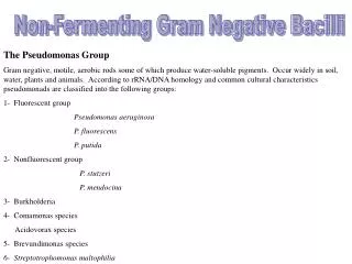
Pseudomonas aeruginosa – General characteristics • Obligate aerobe • small gram-negative rods with a single polar flagellum, produce oxidase & catalase • common inhabitant of soil & water (ubiquitous-wide spread) • grapelike odor • greenish-blue pigment (pyocyanin) • resistant to soaps, dyes, quaternary ammonium disinfectants, drugs, drying • frequent contaminant of ventilators, IV solutions, anesthesia equipment • opportunistic pathogen • multidrug resistant Dr Ekta Page 29
Pathogenicity • common cause of nosocomial infections in hosts with burns, neoplastic disease, cystic fibrosis • Can cause: pneumonia, UTI, abscesses • Septicemia can lead to: endocarditis, meningitis, bronchopneumonia • Corneal ulcers from contaminated lens solutions • Ear infections (Otitis) “swimmer’s ear” • Skin rash (contaminated hot tubs, saunas, swimming pools) Dr Ekta Page 30

![GRAM NEGATIVE BACILLI- MICRO {ST1]](https://cdn1.slideserve.com/2240310/gram-negative-bacilli-micro-st1-dt.jpg)