Download

1 / 13
130 likes | 167 Vues
Delve into the intricate structures and functions of the integumentary system, focusing on the epidermis and dermis, as well as the hypodermis. Learn about the diverse roles of the skin, from regulating body temperature to providing immunity. Explore the different cell types and layers of the epidermis, along with the fascinating world of skin pigments like melanin. Enhance your understanding of skin histology and its significance in overall health and well-being.

E N D
The Integumentary System Chapter 5
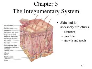
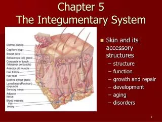
The Skin • epithelial and connective tissues working together • the largest organ of the body • 1.5 - 2 square meters • 4 - 5 kg • variable thickness: 0.5 mm to 4 mm
The Architecture of the Skin 2 main parts: • Epidermis – keratinized stratified squamous epithelium • Dermis – areolar & dense irregular connective tissues Beneath the dermis: • Hypodermis (the subcutaneous layer) – separates skin from muscle; contains areolar and adipose tissues
Skin’s Many Functions • regulation of body temperature • protection – a physical barrier & water conservation • sensation – due to sensory nerve endings • excretion – sweat • immunity – epidermis contains phagocytes • synthesis of vitamin D – for calcium absorption
The Epidermis 4 cell types: • Keratinocytes filled with protein keratin; waterproof barrier • Melanocytes produce pigment melanin • Langerhans cells phagocytes (function in immunity); easily damaged by UV light • Merkel cells detect touch sensations
Epidermal Cell Layers • Stratum basale • a single layer; mitosis pushes the other layers to the top; Merkel cells & melanocytes • Stratum spinosum • 8 to 10 layers of closely packed cells; Langerhans’ cells • Stratum granulosum • 3-5 layers of flattened non-dividing cells; produce large amount of keratin; nuclei & organelles disintegrate
Epidermal Layers Cont. • Stratum lucidum • only in thick skin • 3-5 layers of clear, flat dead cells with keratin • Stratum corneum • 25-30 layers of flattened, dead, keratin-filled cells • continuously shed and replaced It takes 2-4 weeks for each cell to move from the stratum basale to stratum corneum
Epidermal Histology • Stratum Corneum • Stratum Granulosum • Stratum Spinosum • Stratum Basale
Skin Pigments • Hemoglobin – red, carries oxygen in red blood cells • Carotene – yellow/orange,converted to vitamin A, used in the synthesis of vision pigments • Melanin – yellow/red or brown/black
Melanin Cont. • The number of melanocytes is similar in all races – but the amount of melanin produced varies • The UV ↑ production of melanin; melanin protects the body against UV radiation by absorbing UV • Albinism - inability to produce melanin; genetic