Chapter 5 Integumentary System
Chapter 5 Integumentary System. Lecture 5a - Skin Structure. Integumentary system. Includes the skin, sweat and oil glands, hairs, and nails Major Function – protection Accounts for about 7% of total body weight in the average adult (9-11 pounds). Skin Structure. Figure 5.1.


Chapter 5 Integumentary System
E N D
Presentation Transcript
Chapter 5Integumentary System Lecture 5a - Skin Structure
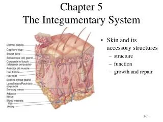
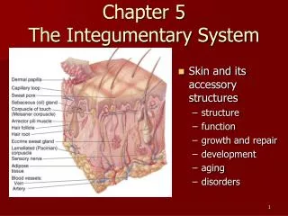
Integumentary system • Includes the skin, sweat and oil glands, hairs, and nails • Major Function – protection • Accounts for about 7% of total body weight in the average adult • (9-11 pounds)
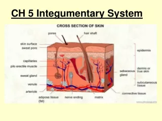
Skin Structure Figure 5.1
Skin Structure • Two layers • 1. Epidermis - outermost layer • Epi = upon • Stratified squamous epithelium • Often keratinized (hardened by keratin)
Skin Structure • Two layers • 2. Dermis • Dense connective tissue • Only Dermis is vascularized • Nutrients reach the epidermis by diffusion
Hypodermis • Subcutaneous tissue • Just deep to skin • Not considered part of skin • Functions as a shock absorber and insulates - Prevents heat loss from body • Mostly adipose tissue (so it stores fat)
Hypodermis • The hypodermis thickens during weight gain • In females, the hypodermis first thickens in thighs and breasts
Hypodermis • In males, the hypodermis first thickens in the anterior abdomen - Sometimes described as a ‘beer belly’
Hypodermis • Loosely anchors skin to underlying structures (mostly muscles) • Skin is loose enough to slide somewhat over structures – many blows glance off our bodies.
Epidermis • Composed of keratinized stratified squamous epithelium • Consists of four distinct cell types and four or five layers • Outer portion of the skin is exposed to the external environment and functions in protection
Cells of the Epidermis • Most are Keratinocytes – produce the fibrous protein keratin • Keratin gives the epidermis protective properties • Kera = horn in Greek • Connected by desmosomes • Keratinocytes arise from the deepest layer of epidermis whose cells undergo continual mitosis
Cells are pushed upward by the production of new cells underneath • When keratinocytes reach surface they are dead, scalelike structures filled with keratin
Millions of these dead cells rub off daily • We have a totally new epidermis every 25 to 45 days • Persistent friction causes accelerated cell and keratin production and a thickening of the epidermis called a callus
Melanocytes – spider-shaped cells that produce the brown pigment melanin (Melan = black)
As melanin is made it accumulates in the processes (the arms) of the melanocytes cells. • Then they are taken up by the keratinocytes and accumulate on the superficial side of the keratinocyte nucleus • Forms a pigment shield to protect nucleus from ultraviolet radiation in sunlight
Other Cells of the Epidermis • Langerhans’ cells – star-shaped cells that arise from bone marrow and migrate to the epidermis as epidermal macrophages that help activate the immune system • Merkel cells – function as touch receptors in association with sensory nerve endings
Layers of the Epidermis Figure 5.2b
Layers of the Epidermis • Thick skin • 5 layers or strata (strata = sheets) • Covers palms, fingertips, soles of feet • Thin skin • 4 layers
Layers of the Epidermis: Stratum Basale (Basal Layer) • Deepest epidermal layer firmly attached to the dermis • It is a single row consisting of mostly young keratinocytes, with 10-25% melanocytes, and an occasional Merkel cell • Cells undergo rapid division, (mitosis) hence its alternate name, stratum germinativum
Layers of the Epidermis: Stratum Spinosum (Prickly Layer) • Several cell layers thick • Cells contain a weblike system of intermediate filaments attached to desmosomes • Melanin granules and Langerhans’ cells are abundant in this layer
Layers of the Epidermis: Stratum Granulosum (Granular Layer) • Thin; three to five cell layers in which drastic changes in keratinocyte appearance occurs • Cells flatten, nuclei & organelles disintegrate, and keratohyaline and lamellated granules accumulate
Layers of the Epidermis: Stratum Basale (Basal Layer) Figure 5.2b
Layers of the Epidermis: Stratum Lucidum (Clear Layer) • Present only in thick skin • Thin, transparent band superficial to the stratum granulosum • Consists of a few rows of flat, dead keratinocytes
Layers of the Epidermis: Stratum Corneum • Outermost layer of keratinized cells • Accounts for three quarters of the epidermal thickness • Functions include: • Waterproofing • Protection from abrasion and penetration • Rendering the body relatively insensitive to biological, chemical, and physical assaults
Average person sheds 40 pounds of skin flakes from the stratum corneum layer in a lifetime • Provides food for dust mites!
Dermis • Second major skin region containing strong, flexible connective tissue • Cell types include fibroblasts, macrophages, and occasionally mast cells and white blood cells • Composed of two layers – papillary and reticular
Dermis • Richly supplied with nerve fibers, blood vessels, and lymphatic vessels. • Most hair follicles, oil and sweat glands are derived from epidermal tissue but reside in the dermis • In animals, this is the hide that makes leather products
Layers of the Dermis: Papillary Layer • Areolar connective tissue with collagen and elastic fibers and blood vessels
Layers of the Dermis: Papillary Layer • Its superior surface contains peglike projections called dermal papillae
Layers of the Dermis: Papillary Layer • Dermal papillae contain capillary loops, touch receptors called Meissner’s corpuscles, and free nerve endings
On palms of the hands and soles of the feet, dermal papillae lie on top larger mounds called dermal ridges • The epidermis that lies on the dermal ridges is called the epidermal ridges • These epidermal ridges are genetically determined and unique to each of us • These are our fingerprints!
Layers of the Dermis: Reticular Layer • Accounts for approximately 80% of the thickness of the dermis • Is dense irregular connective tissue • Collagen fibers in this layer add strength and resiliency to the skin • Elastin fibers provide stretch-recoil properties
Most bundles of collagen fibers run parallel to the skin surface. • Less dense regions of collagen fibers form cleavage or tension lines in the skin – invisible from surface • These cleavage lines are important to surgeons • Incisions made parallel to these lines, the skin gapes less and heals more readily than when the incision is made across cleavage lines
Epidermal ridges (fingerprints) and cleavage lines are called skin markings • Another type of skin marking is Flexure lines • These are externally visible • Dermal folds that occur at or near joints • See them as creases in your palms, wrists, finges, soles, and toes
Homeostatic imbalances • Stretching of the skin can tear the dermis • Indicated by silvery white scars called striae (streaks) commonly called stretch marks
Homeostatic imbalances • Stretching orTrauma like a burn or a poor fitting shoe, or digging a hole with a shovel, can cause a blister • Separation of the epidermal and dermal layers by a fluid filled pocket
Skin Color • Three pigments contribute to skin color • Melanin – yellow to reddish-brown to black pigment, responsible for dark skin colors • Freckles and pigmented moles – result from local accumulations of melanin • Carotene – yellow to orange pigment, most obvious in the palms and soles of the feet • Hemoglobin – reddish pigment responsible for the pinkish hue of the skin
Melanin • Made in melanocytes and passed to keratinocytes • All humans have about the same number of melanocytes • Individual differences in skin coloring reflect the amount of melanin made and retained • Darker skinned people produce more melanin and their keratinocytes retain it longer
Melanin • Melanocytes are more active when exposed to sunlight • Protects DNA of skin cells from UV radiation by absorbing the light and dissipating the energy as heat • Causes a ‘tan’
Even with Melanin’s protection, excess sun exposure eventually damages the skin • Leathery skin, depressed immune system, skin cancer
Carotene and hemoglobin • Carotene – yellow to orange pigment (also found in carrots) • Accumulates in the stratum corneum and the hypodermis • Most intense when carotene-rich foods are eaten • Hemoglobin – pinkish hue reflecting the crimson color of oxygenated hemoglobin in red blood cells in the dermal capillaries
Homeostatic imbalances • When hemoglobin is poorly oxygenated, both the blood and the skin appear blue • Called cyanosis (cyan = dark blue) • Lots of melanin may mask cyanotic appearance so dark-skinned individuals will only show blue tint in their mucous membranes and nail beds
Homeostatic imbalances • Skin often becomes cyanotic during heart failure and severe respiratory disorders
Homeostatic imbalances • Alterations in skin color signal certain diseases and emotional stimuli • Redness – may indicate embarrassment, fever, hypertension, inflammation, or allergy
Homeostatic imbalances • Jaundice or yellow cast – usually signifies a liver disorder
Homeostatic imbalances • Bronzing – a bronze or almost metallic appearance of the skin is a sign of Addison’s disease • Black and Blue marks or bruises – reveal where blood escaped from the circulatory system and clotted beneath the skin – called hematomas