Download
1 / 87
870 likes | 895 Vues
CH 5 Integumentary System. SKIN. The integumentary system includes the skin and its accessory organs Another term for the skin is the “cutaneous membrane”. Facts about the SKIN…. Skin is waterproof The outer surface of the skin is made of dead cells. House dust is mainly skin flakes!

E N D
SKIN • The integumentary system includes the skin and its accessory organs • Another term for the skin is the “cutaneous membrane”.
Facts about the SKIN… • Skin is waterproof • The outer surface of the skin is made of dead cells. House dust is mainly skin flakes! • If you laid out all your skin on a flat surface, it would have an area of about 2 square meters (22 square feet). Skin weighs about 2.5 kilograms (12-15% of body weight) - the largest organ in the body. • What hurts if you pull it, but doesn't hurt if you cut it? • Your hair, of course! • Skin is elastic - it springs back into shape when stretched.
Skin Facts Continued…. • Some medicines (eg: oestrogen, nicotine) can pass through the skin, but others cannot (eg: insulin). Why is that? Because only fat-soluble substances can enter the skin, not water-soluble ones. • Your hair stands on end and you develop goose bumps because there are tiny muscles attached to the hair follicles and they contract when you are frightened or cold.
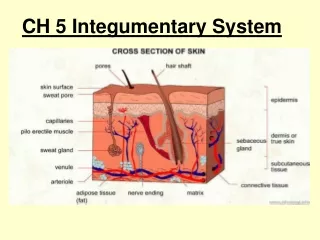
The Skin and its Tissues • Skin functions: • Protective covering (primary function) – barrier to the entry of microbes, viruses, and UV exposure • Aids in regulating body temperature • Slows water loss • Houses sensory receptors • Synthesizes various chemicals (vitamin D and melanin) • Excretes waste • The skin is composed of an epidermis, dermis, and subcutaneous layer (hypodermis).
THE SKIN Epidermis Dermis Subcutaneous (Hypodermis)
The EPIDERMIS • 4-5 layers of 4 different types of cells. • The deepest layer of the epidermis contains cells undergoing cell division (mitosis) to produce new cells. • About 90% of the epidermis are keratinocytes. • Epidermal cells become keratinocytes when they undergo keratinization. During this process, they mature and are pushed toward the surface while producing keratin (takes about 2-4 weeks). What does keratin do? • Gives tissue a waterproof quality • Melanocytes produce the skin pigment melanin. Melanin is a brown/black pigment that covers the nucleus to protect the DNA from UV radiation- which can cause mutation. The more that cells are exposed to UV radiation, the more melanin is produced. A sun tan is a sign of injury!!
Epidermis Continued…. • Langerhans cells- Arise from red bone marrow and move to the epidermis where they participate in immune responses. • Merkel cells- Found in the deepest layer of the epidermis where they contact the flattened part of a sensory nerve cell and function in the sensation of touch.
Layers of the Epidermis- from the bottom to top • Stratum Basale- Here is where cell division takes place, producing new skin cells and pushing older cells toward the surface. This layer contains pre-keratinocytes, melanocytes, Langerhans cells and Merkel cells. This layer is attached to a layer of fibers called the basement membrane. • Stratum Spinosum- Contains spiny keratinocytes that are not yet mature enough to produce keratin. • Stratum Granulosum- Contains keratin producing keratinocytes. • Stratum Lucidum- Clear, flat, dead cells found only in the thick skin of palms and soles. • Stratum Corneum- Thickest layer- 30 layers of flat, dead keratinocytes.
Quick Review • Another name for skin is the ______________ membrane. • What are 5 functions of skin? • What are the 3 layers of skin? • What are the 4 types of cells found in the epidermis? • What are the 5 layers of the epidermis? • Which layer contains functioning keratinocytes? • Which layer is attached to the basement membrane? • Which layer is the thickest?
The DERMIS • Composed of 2 layers: papillary layer and reticular layer. • Papillary layer contains a thin arrangement of collagen fibers and supplies nutrients to select layers of the epidermis and regulates temperature (by increasing and reducing blood supply to the epidermis). The fingerlike projections of this layer are called dermalpapillae. These cause ridges in the epidermis and are what produce fingerprints. • Inside the dermal papillae are blood vessels and nerve endings. The nerve endings are called corpuscles of touch or Meissner corpuscles. These are sensitive to touch. What type of epidermal cell is closely associated with these? • Merkel Cells!
Dermis continued….. • Reticular layer- The reticular layer contains thicker collagen fibers than the papillary dermis. It strengthens the skin, provides structure and provides elasticity. It also supports other components of the skin, such as hair follicles, nerves, oil glands, muscles, and sweat glands.
HAIR • Also called pili- Each strand is dead, keratinized cells that consist of a shaft (above the skin) and a root (below the skin). • Surrounding the root is a hair follicle which includes an external root sheath, internal root sheath, and connective tissue sheath • The base of the follicle is the matrix- where new hair cells are formed from cell division • Surrounding the follicle base is/are: • blood vessels- provide nourishment to the hair • arrector pili muscle-contracts and causes the hair to stand up (goose bumps) • hair root plexus (nerve endings)
Sebaceous Glands • Sebaceous glands usually are associated with hair follicles • Secrete sebum, which keeps hair from drying out • If plugged and infected…a pimple develops
Sweat (Sudoriferous) Glands • Each sweat gland consists of a coiled tube (duct) • 2 Types (Apocrineand Eccrine) • Apocrine glands respond to emotional stress – larger and occur in armpits (axillary regions) and groin areas…these produce a solution that bacteria act on to produce “body odor” • Eccrine glands respond to elevated body temperature • Sweat is primarily water, but also contains salts and wastes
The SUBCUTANEOUS Layer (Hypodermis) • Adipose (fat) tissue helps conserve body heat • Contains blood vessels that branch into the dermis • The layer where you receive shots and vaccinations…
Nails • Nails are produced by epidermal cells originating at the nail matrix that undergo keratinization HOW SWEET ARE THOSE NAILS??
Skin Cancer Facts Most skin tumors are benign (non-cancerous)– ex. Warts, calluses, moles Skin cancers metastasize, which means that they invade other tissues. If they enter the bloodstream and/or lymph system, they can travel anywhere and lodge themselves in other locations. The cause of skin cancer is not known, but overexposure to ultraviolet (UV) radiation is the main risk factor. The DNA in a cell can become mutated, and consequently, it divides out of control.
Basal Cell Carcinoma Least malignant and most common skin cancer Most often appears on the exposed areas of the face Appears as a shiny dome-shaped nodule that later develops a central ulcer with a “pearly” beaded edge Full cure in 99% that are removed surgically Cells of the stratum basale no longer form keratin and do not honor the boundary between dermis and epidermis
Squamous Cell Carcinoma Arises from the cells of the stratum spinosum Occurs most often on the scalp, ears, hands, and lower lip Appears as a scaly, reddened elevation that gradually forms a shallow ulcer with a firm, raised border It grows rapidly and metastasizes to adjacent lymph nodes if not removed Chance for complete cure is good if caught early and removed surgically or by radiation therapy
Malignant Melanoma Cancer of the melanocytes 5% of all skin cancers Is often deadly and chance of survival is 50%...early detection helps. Can begin anywhere there is pigmentation, some develop from pigmented moles. Usually appears as a spreading brown to black patch that metastasizes rapidly to surrounding lymph nodes and blood vessels. Use the ABCDE Rule to recognize it
The ABCDE rule is a convenient guide to the usual signs of melanoma. A is for ASYMMETRY: One-half of a mole or birthmark does not match the other. B is for BORDER: The edges are irregular, ragged, notched, or blurred. C is for COLOR The color is not the same all over, but may have differing shades of brown or black, sometimes with patches of red, white, or blue. D is for DIAMETER: The area is larger than 6 millimeters (about ¼ inch -- the size of a pencil eraser) E is for EVOLVING: If the growth changes at all
IMPORTANT NOTE: One blistering sunburn in childhood or adolescence more than doubles a person's chances of developing melanoma later in life. A person's risk for melanoma also doubles if he or she has had five or more sunburns at any age.
Burns • Serious threat to skin • 2 Life Threatening Problems associated with Burns: • Body Fluid Loss-. Dehydration and electrolyte imbalance follow and can lead to shutdown of kidneys and circulatory shock • Infection- The leading cause of death in burn victims. Burned skin is sterile for about 24 hrs. The bacteria and fungi easily invade areas where the skin is destroyed and feed off of the dead tissues. The patient’s immune system becomes overwhelmed and suppressed after severe burn injury.
Rule of Nines • The rule of nines assesses the percentage of a person’s body that is burned and is used to help guide treatment decisions. • This divides the body into regions and states the surface area % of the body for each region. See next slide.