Understanding the Skin: An In-depth Guide
240 likes | 354 Vues
Delve into the intricate layers and functions of the integumentary system, including the epidermis, dermis, hair follicles, glands, and more. Learn about the roles of cells like keratinocytes and melanocytes, as well as the vital function of blood vessels and subcutaneous tissue.

Understanding the Skin: An In-depth Guide
E N D
Presentation Transcript
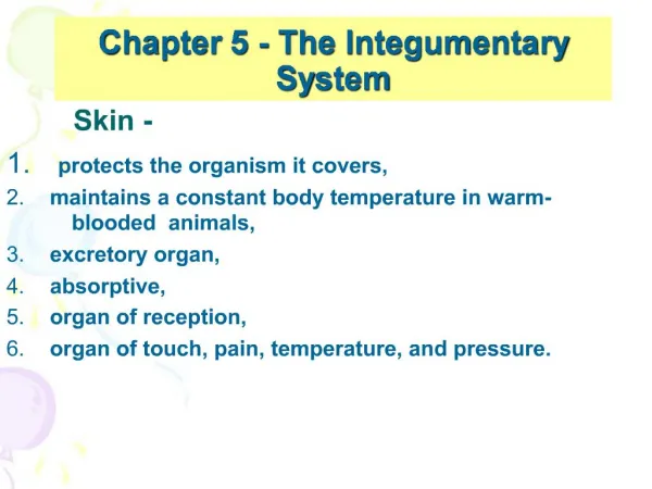
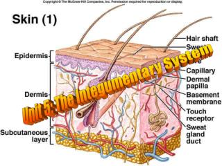
Introduction • Integument consists of: • Skin – largest organ • Accessory structures (hair/sweat glands) • Subcutaneous tissue • Skin • Barrier to many harmful substances
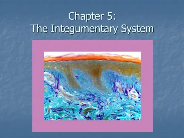
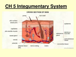
The Skin • 2 major layers (Fig. 5-1) • Epidermis – outer layer • Dermis – inner layer
Epidermis • Stratified squamous keratinizing epithelium • Thickest on palms, soles • Abundant keratinocytes • No capillaries • 2 sublayers • inner stratum germinativum • outer stratum corneum
Stratum Germinativum • “stratum basale” • Base of the epidermis where mitosis occurs • older cells pushed toward skin surface • produce keratin and die • Merkel cells (Merkel discs) • touch receptors (Fig. 5-2)
Stratum Germinativum • Keratinocytes • living cells that synthesize antimicrobial “defensins” • rupture pathogen membranes as part of inflammatory process • Living portion produces vitamin D when exposed to sunlight
Stratum Corneum • Outermost epidermal layer • Keratin prevents evaporation and water entry • Barrier to pathogens & chemicals
Langerhans Cells • “dendritic cells” (Fig. 5-2) • Originate in red bone marrow • Phagocytize foreign material • migrate to lymph nodes; take pathogen to lymphocytes • triggers immune response
Melanocytes • See Fig. 5-2 • Produce melanin • those with darker skin produce large amounts • production increased in when exposed to UV rays • Melanin gives color to hair, iris, choroid layer of eye • See Table 5-1
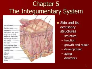
Dermis • Made of irregular fibrous connective tissue • fibroblasts produce collagen & elastic fibers • allow strength & elasticity • Papillary layer – junction of dermis with epidermis (Fig. 5-1) • abundant capillaries • Accessory structures • hair & nail follicles, sensory receptors, glands
Hair Follicles • Made of epidermal tissue • Base of follicle (Fig. 5-3) • Hair root; mitosis occurs in matrix • Produce keratin • Get color from melanin • Die & become incorporated into hair shaft • Hair shaft pushed toward skin surface
Hair Follicles • Eyelashes & eyebrows keep dust & sweat out of eyes • Nostril hairs keep dust from entering nasal cavities • Hair on head provides insulation • body hair doesn’t serve this purpose • Pilomotor (arrectorpili muscle) attached to each follicle • pull hair follicles upright (fear, cold)
Nail Follicles • On ends of fingers & toes • Produce nails; mitosis in nail root at the nail’s base (Fig. 5-4) • New cells produce keratin & die • Nail is dead keratin cells, but nail bed is alive
Nails • Protect fingers & toes from mechanical injury • Allow dexterity • Good for scratching
Receptors • Cutaneous senses • touch, pressure, heat, cold, pain • Specific receptor for each sensation • Receptors & sensation provide CNS with information about external environment
Glands • Made of epithelial tissue (Fig. 5-1) • Sebaceous glands • Secrete sebum (oil) • inhibits bacterial growth on skin’s surface • prevents drying of skin, hair • Ceruminous glands • in the dermis of ear canals • secrete cerumen (ear wax) • keeps outer surface of eardrum pliable; prevents drying
Glands • Sweat glands • 2 types • Apocrine – numerous in axillae (underarm), genital areas • Eccrine – numerous on forehead, upper lip, palms, soles
Blood Vessels • Capillaries in dermis • Arterioles • Smooth muscle in walls permits constriction or dilation • maintains body temp.
Subcutaneous Tissue • “superficial fascia” • Made of areolar and adipose tissue • Connects dermis to underlying muscles • Secondary line of defense
Aging & The Integumentary System • Skin layers become thinner, more fragile • Fibroblasts die & are not replaced • Repair of breaks or cuts slows • Skin wrinkles • Glands become less active • Skin becomes dry • Temp. regulation becomes harder • Hair thins, turns white