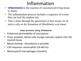
Inflammatory Patterns and Exudates in Acute Inflammation
350 likes | 472 Vues
Explore the various morphological patterns of inflammation, including serous, catarrhal, fibrinous, hemorrhagic, suppurative, pseudomembranous, ulcerative, and gangrenous. Learn about different types of exudates, cellular participants in host defense, and the distinction between acute and chronic inflammation.

Inflammatory Patterns and Exudates in Acute Inflammation
E N D
Presentation Transcript
Serous Catarrhal Fibrinous Hemorrhagic Suppurative Pseudomembranous Ulcerative Gangrenous PATTERNS ACUTE INFLAMMATION
INFLAMMATORY EXUDATES • Serous • Watery, protein-poor effusion (e.g., blister) • Serous – largely plasma, low in protein, Occurs early or in mild inflammation
ACUTE INFLAMMATION SEROUS
ACUTE INFLAMMATION SEROUS
INFLAMMATORY EXUDATES • Catarrhal – mucus hypersecretion that accompanies inflammation of a mucus membrane.
ACUTE INFLAMMATION CATARRHAL
INFLAMMATORY EXUDATES • Fibrinous – large amounts of fibrinogen, Forms a thick, sticky meshwork. Only removed by fibrolytic enzymes. Failure of removal leads to influx of fibroblasts & scar tissue formation
ACUTE INFLAMMATION FIBRINOUS
INFLAMMATORY EXUDATES • Hemorrhagic – damage to blood vessels, Occurs with other forms of exudate.
ACUTE INFLAMMATION HEMORRHAGIC
INFLAMMATORY EXUDATES • Suppurative/ purulent • Presence of pus (pyogenic staph spp.) • Often walled-off if persistent – contains pus (remains of WBCs, protein and tissue debris). Liquefactive necrosis!
ACUTE INFLAMMATION SUPPURATIVE / PURULENT - ABSCESS
ACUTE INFLAMMATION SUPPURATIVE / PURULENT - ABSCESS
ACUTE INFLAMMATION SUPPURATIVE / PURULENT - EMPYEMA
ACUTE INFLAMMATION SUPPURATIVE / PURULENT
Pseudomembranous • Adherent layer of inflammatory cells & debris at the site of mucosal injury • Pseudomembranous colitis – Clostridium difficile • Diphtheria – Corynebacterium diphtheriae
ACUTE INFLAMMATION PSEUDOMEMBRANOUS Atlanta South Gastroenterology, P.C.
Ulceration & erosion • Local defects or excavations of the surface of an organ or mucous membrane resulting from sloughing or loss of necrotic tissue. An ulcer is full thickness epithelial loss (through basement membrane). An erosion is more superficial and does not penetrate basement membrane.
ACUTE INFLAMMATION ULCERATIVE
ACUTE INFLAMMATION GANGRENOUS
ACUTE INFLAMMATION GANGRENOUS Appendix Gallbladder
HOST DEFENSE POLYMORPHONUCLEAR LEUKOCYTES (PMNL) Neutrophils Eosinophils Basophils
HOST DEFENSE MONONUCLEAR LEUKOCYTES Lymphocytes Monocytes
Phagocytes • Derived from the Greek words “Eat & cell”. • Phagocytosis is carried out by macrophages, neutrophils
Neutrophil - common leucocyte of the blood - 40-70% - short-lived phagocytic cell - predominate early in infection - ACUTE INFLAMMATION
Monocyte- largest nucleated cell of blood - 2-10% -develops into macrophage when it migrates to tissues Macrophage- phagocyte--scavenger cell-- of tissues - CHRONIC INFLAMMATION
Functions of macrophages • Phagocytosis • Antigen presentation • Cytokines
EOSINOPHIL • 1-6% in peripheral blood • Allergic reactions • Parasitic infestations • Release mediators
BASOPHILS & MAST CELLS • Basophils in blood - 0-1% • Mast cells – in tissues • IgE surface receptor • Allergic reactions