11262 2012 808 MOESM2 ESM
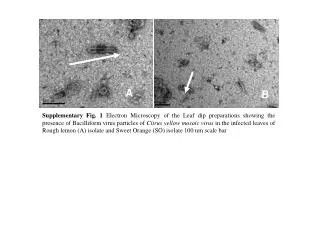
A. B. Supplementary Fig. 1 Electron Microscopy of the Leaf dip preparations showing the presence of Bacilliform virus particles of Citrus yellow mosaic virus in the infected leaves of Rough lemon (A) isolate and Sweet Orange (SO) isolate 100 nm scale bar.

11262 2012 808 MOESM2 ESM
E N D
Presentation Transcript
A B Supplementary Fig. 1 Electron Microscopy of the Leaf dip preparations showing the presence of Bacilliform virus particles of Citrus yellow mosaic virus in the infected leaves of Rough lemon (A) isolate and Sweet Orange (SO) isolate 100 nm scale bar
Supplementary Fig. 2 Alignment of CMBVSOJNTU, CMBVROL ORF II protein with CSSV ORF II Product at secondary structural level Cylinder region – Helix region; Solid Arrow – Sheet; Solid line – Loop region
Supplementary Fig. 3 Alignment of CMBV ORF III encoded protein sequences with RTBV Coat Protein showing presence of conserved pockets and also the presence of a Cysteine rich motif (Zinc finger like domain represented in a box) at the C-terminus of the putative coat protein domain
Supplementary Fig. 4 Alignment of CMBVSOJNTU, CMBVROL ORF III protein with RTBV Coat Protein (ORF III Product) at secondary structural level Cylinder region – Helix region; Solid Arrow – Sheet; Solid line – Loop region