Three-dimensional hydrogels for biomedical applications
360 likes | 753 Vues
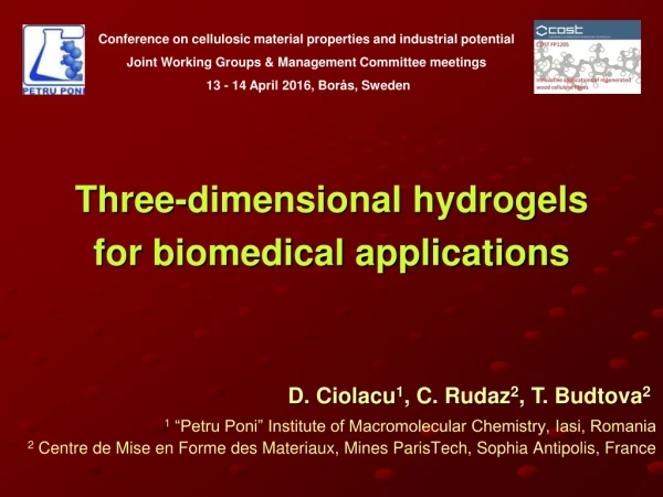
Conference on cellulosic material properties and industrial potential Joint Working Groups & Management Committee meetings 13 - 14 April 2016, Borås, Sweden. Three-dimensional hydrogels for biomedical applications. 1 “Petru Poni” Institute of Macromolecular Chemistry, Iasi, Romania

Three-dimensional hydrogels for biomedical applications
E N D
Presentation Transcript
Conference on cellulosic material properties and industrial potential Joint Working Groups & Management Committee meetings 13 - 14 April 2016, Borås, Sweden Three-dimensional hydrogels for biomedical applications 1 “Petru Poni” Institute of Macromolecular Chemistry, Iasi, Romania 2 Centre de Mise en Forme des Materiaux, Mines ParisTech, Sophia Antipolis, France D. Ciolacu1, C. Rudaz2, T. Budtova2
Background • Cellulose and cellulose derivatives - based hydrogels are very attractive materials for bio-medical applications due to their biocompatibility and biodegradability. • “Cellulose hydrogel” is a term which is historically used either for cellulose coagulated (or regenerated) or for cellulose nanofibresdispersed in water (either bacterial or nanofibrillated or cellulose whiskers). • Cellulose II - based hydrogels are obtained as an inevitable step during cellulose processing, i.e. cellulose coagulation into water from any solution. • Cellulose hydrogel can be prepared by coagulation or regeneration of cellulose from various cellulose solutions: NMMO monohydrate, LiCl/DMAc, ionic liquids and 6-10 wt% NaOH-water based solvent.
Objectives • to investigate the properties of cellulose II - based cryogels prepared from cellulose - 8% NaOH-water solutions • to investigate the influence of ECH concentration on swelling of hydrogels • to demonstrate their possible application as delivery device by incorporating procaine hydrochloride (PrHy)
Physical gelation coagulation freeze - drying Cellulose - Cellulose gel Cellulose Cellulose 8%NaOH - water hydrogel cryogel solution PrHy Chemical gelation ECH Preparation of cellulose II - basedcryogels Schematic presentation of physically and chemically cross-linked cellulose cryogels loaded with PrHy.
8%NaOH-water ( physical gelation) ECH (chemical gelation) Materials • Relative concentration of ECHwasvaried to reachdifferent • molar ratio of ECH to AGU (Rrel = 0, 0.25, 0.5, 1, 2 and 4) • 5 and 7 wt% cellulose concentrations
What is the effect of cross-linking on cellulose structure and crystallinity ?
Structural properties of cryogels - solid-state 13C NMR Chemical cross-linking leads to spectra modifications • with the increase of ECH conc. • a loss in the resolution of C4and C6 • the peak corresponding to C2,3,5is broadened - lower mobility of carbons on AGU due to the cross-linking of cellulose with ECH
Structural properties of cryogels - solid-state 13C NMR Normalised areas of peaks C2,3,5 and C6 as a function of molar ratio Rrel • the surface area under C6peak is independent on Rrel, • the surface under C2,3,5 increases with Rrel - in the cross-linking reaction of cellulose with ECH the most reactive hydroxyl groups are C2
Structural properties of cryogels - XRD • Physical cross-linked cryogels – cellulose II difractogram • Chemical cross-linked cryogels – amorphous cellulose difractogram X-ray diffractograms of cellulose cryogels: 5%R0, 5%R1 and 5%R2
Structural properties of cryogels - XRD X-ray diffractograms of cellulose cryogels: 7%R0, 7%R0.5 and 7%R1
Structural properties of cryogels - DSC • conditioned samples • RH: 65 % • temp: 25 oC, the enthalpy increases with the increase of ECH concentration (Rrel) • the increase of the amorphous fraction of cellulose
Structural properties of cryogels – NMR, XRD,DSC Crystallinity indexes and dehydration heat (ΔH) for cellulose cryogels • NMR method gives lower crystallinities than the XRD due to the fact that the C in the amorphous region have a chemical shift distinct from C in the crystalline region and XRD does not distinguish cellulose chains on the surface of cellulose crystallites, as NMR does.
What is the effect of cross-linking on swelling of gels and cryogels in water?
Swelling of gels and cryogels Two types of swelling experiments were performed: hydrogels obtained after cellulose complete coagulation (never dried, at equilibrium), cellulose cryogels swelling in water, at 37 °C. • the absolute values of cryogel swelling are lower– cryogels (900 - 2500 %), hydrogels (1300 - 3500 %) - explained by the well-known hornification phenomenon. • the “anomalous” swelling, i.e. increase in swelling degree with the increase of ECH concentration up to a certain value, is observed even for freeze-dried samples.
DVS measurements of cryogels Sorption/desorption isotherms of cryogels • The isotherms of the cellulose-based cryogels show hysteresis at RH > 60%, this being usually attributed to pore effects. • An increase of the cross-linking degree of the cryogels determined an increase in the maximum water vapor sorption capacity.
Density of cellulose cryogel • The bulk density decreases with the increase of ECH until Rrel> 1, as expected because of gels swelling during coagulation. • The bulk density for the physically cross-linked cellulose is twice higher (5% R0 - 0.108 g/cm3) than that of chemically cross-linked sample, (5% R1 - 0.042 g/cm3).
SEM observations • physically cross-linked cryogel – heterogeneous morphology. • chemically cross-linked cryogel – increased pore size, the morphology is foam-like and it disappear the denser regions. 5% R0 5% R1 5% R2 5% R4
Kinetics of cellulose cryogel swelling Evolution of the reduced swelling degree as a function of time, at 37 °C, for cellulose cryogels • All swelling data fall together, thus the swelling rate constant, kswdoes not depend either on cellulose concentration or type of cross-linking. • The reason is that cellulose concentration in cryogels is very low and pores are very large; thus, the diffusion of water is not sensitive to the variations in cellulose cryogels morphology.
Release profiles of PrHy from cryogels Release profiles of PrHy in water at 37 °C from: (a) cellulose cryogels 5% R0 loaded with different amount of PrHy; (b) cellulose cryogels loaded with 4 g/L PrHy at different Rrel • The release is completed in about 3 h, much slower than cryogel swelling in water (~ 1h). • As expected, higher the initial drug concentration, higher amount of drug is released. • Higher swelling of cellulose leads to more uptake of drug during loading.
Release profiles of PrHy from cryogels Release profiles of PrHy in water at 37 °C from cryogels prepared from 5 wt% solutions as a function of Rrel. • The rate of release is higher for chemically cross-linked cellulose compared to their physically cross-linked counterparts → lower density and larger pores of cryogels. • Procaine is an asymmetric molecule from of amino-ester group. Thus, the rate of PrHy release is one order of magnitude lower that the rate of cryogels swelling in water.
Schematic presentation of the structure of physical and chemical cross-linked cellulose gels
Summarised experimental results • The experimental results obtained on the structure and properties of physically and chemically gelled cellulose: • NMR, XRD and DSC show that in the presence of ECH cellulose crystallinity decreases compared to cellulose II obtained via physical gelation. • The swelling of cellulose gel (never-dried, during coagulation) and of dry cellulose cryogels in water increases with the increase of ECH concentration, in the region of [ECH] < 2. • Humidity adsorption by cellulose cryogels increases in the presence of ECH. • Density of chemically cross-linked cellulose cryogels is lower than that of their physically gelled couterparts.
Network formation in cellulose solutions • Physical gelation occurs due to cellulose chains self-association because of the preferential cellulose-cellulose and not cellulose-solvent interactions; in addition, physical gelation is accompanied by a micro-phase separation (gels become opaque). • Chemical gelation i) perturbs cellulose chains self-association → chemical bonds act as “spacers” and leads to the decrease of crystallinity; ii) leads to a more homogeneous structure which results in transparent swollen coagulated cellulose hydrogels; iii) increases swelling in water due to a more porous structure.
Conclusions • Cellulose cryogels were prepared from cellulose-8%NaOH-water solutions by gelation, coagulation in water followed by freeze-drying. Gelation was either viaphysical (“aging” of cellulose solutions) and chemical (cross-linking with different amounts of ECH). • The results allowed us to suggest a hypothesis: while during physical gelation chains are self-associating forming a heterogeneous network with “thick” walls and pores of various sizes, chemical bonds act as spacers between chains perturbing their self-association and preventing packing. • In chemically cross-linked cellulose larger amount of drug can be loaded and the release kinetics is faster compared to physically cross-linked matrix. • By varying cellulose concentration and amount of cross-linker it is possible to prepare versatile cellulose hydrogels and dry porous networks with controlled morphology and porosity.
Acknowledgements This work was supported by a grant of the Romanian National Authority for Scientific Research and Innovation, CNCS - UEFISCDI, project number PN-II-RU-TE-2014-4-0558. A part of this work was also financial supported by European Union's Seventh Framework Programme (FP7/2007-2013) under grant agreement n°264115 – STREAM and French National Agency for Research (ANR), “Nanocel” project ANR-09-HABISOL-010.