Download
1 / 72
720 likes | 725 Vues
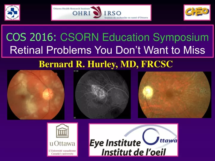
Bernard R. Hurley, MD, FRCSC. COS 2016: CSORN Education Symposium Retinal Problems You Don’t Want to Miss. Financial Disclosure. I have received speaking honoraria from Alcon Novartis Allergan Nikon Bayer I have participated in advisory boards for Bayer Novartis Alcon.

E N D
Bernard R. Hurley, MD, FRCSC COS 2016: CSORN Education Symposium Retinal Problems You Don’t Want to Miss
Financial Disclosure • I have received speaking honoraria from • Alcon • Novartis • Allergan • Nikon • Bayer • I have participated in advisory boards for • Bayer • Novartis • Alcon
Outline – Retinal Problems Not to Miss Outline • Vast subject • Series of 14 interesting cases • Each illustrate a retinal finding with important implications • Common and important • Timely diagnosis and treatment • Important for preserving vision • Rare but serious • Timely diagnosis and treatment • Important for preserving life • Every eye doctor’s dream • Summary
Case #1 : A common scenario • Diagnosis with profound ophthalmic and systemic implications • 73 year old male • Woke up previous morning unable to see anything from right eye
Case #1 : A common scenario • 73 year old male blind in the right eye x 48 hours • IVFA confirms CRAO • NO emboli seen
Case #1: Subsequent Systems Review • Severe jaw pain with eating • “Everything I eat has to go though a blender because I can’t chew anything” • Saw his dentist • TMJ problem, night prosthesis • Severe bi-temporal headaches • Saw his G.P. and given a TENS machine for tension headaches • Transcutaneous electrical nerve stimulation • Low-voltage electrical current for pain relief
Case #1: Subsequent Systems Review • Severe jaw pain with eating • Severe bi-temporal headaches • Shoulder and hip pain • Saw his rheumatologist • Low dose oral prednisone • Polymyalgia rheumatica • Neck pain, weight loss, intermittent blurred vision O.S. • Additional tests ordered to confirm diagnosis
Initial Management • Visual loss caused by giant cell arteritis is a medical emergency • Prompt treatment with systemic corticosteroids • I.V. high dose steroids • First advocated as treatment 25 years ago • A single case report • Currently no consensus regarding the dose, regimen, and duration of treatment • Chan, C.C.K. et al. Steroid management in giant cell arteritis. British Journal of Ophthalmology 2001;85:1061-1064
Treatment Summary Adapted from M. Tariq Bhatti and Homayoun Tabandeh. Giant Cell Arteritis. Current Opinion in Ophthalmology. December 2001;12:393-399.
Case #1 : A common scenario • Diagnosis with profound ophthalmic and systemic implications • GCA • A common clinical scenario • Often seen on formal exams • Keep high index of suspicion • Elderly patient with visual loss • Consider in cases of • CRAO • AION
Case # 2 • 58 year old male • Seen for intermittent blurred vision OU • Abnormal fundus findings prompting retina consult
Very important clinical sign 98% of patients with cotton-wool spots have an associated systemic disease Brown et al. Cotton Wool Spots. Retina 1985;5:206-214 Important Differential: Diabetes HTN Branch/Central Vein Occlusion Ocular Ischemic Syndrome Carotid Emboli Sickle-cell retinopathy Radiation retinopathy Vasculitis (especially Lupus) Leukemia HIV Giant Cell Arteritis Ocular Signs: Cotton-Wool Spots
Case # 2 • 58 year old male • Seen for intermittent blurred vision OU • Abnormal fundus findings prompting retina consult • Good VA OU • ROS • Jaw pain • Headache • Neck stiffness • ESR = 107 • Biopsy • Positive for GCA
Cotton-Wool Spots and Giant Cell Arteritis • Cotton-wool spots in GCA initially reported in 1970 • Advocated as a prominent early ophthalmologic sign • Proceeding irreversible visual loss in GCA • Melberg et al. Cotton-wool spots and the early diagnosis of giant cell arteritis. Ophthalmology 1995; 102(11):1611-4. • “any patient older than 55 with the ophthalmoscopic finding of one cotton-wool spot, even in the absence of other clinical symptoms deserves specific questions regarding constitutional symptoms”
Cases #3 • 60 year old male • Significant cataract OD • Removed with “perfect” surgery • Initial post op period • Excellent improvement in VA • Over next several weeks • Significant reduction in vision • Diagnosed with post op CME • Irvine-Gass syndrome • Intensive topical steroids • No response despite good compliance
Cases #3: CNVM Masquerading as CME • 60 year old male • Expected to see CME • Consider other common diagnosis in the cataract population • Wet AMD • Worsening diabetic retinopathy • Vascular occlusion • Small vein or artery occlusion
Cases #4 • 60 year old male • Significant cataract OS • Removed with “perfect” surgery • Initial post op period • Excellent improvement in VA • Over next several weeks • Significant reduction in vision • Diagnosed with PCO • No improvement with YAG
Cases #4 • 60 year old male • Significant cataract OS • Removed with “perfect” surgery • Dilated Fundus exam: • Central macular cyst • Irvine Gass • Started on PF • Q1H • No response • OCT not typical • Retina opinion • Chronic inferior RD
Case #5: Unexplained Iritis • 60 year old gentleman • Chronic unilaterial iritis • Decreased vision • Unresponsive to steroid therapy • Unusual • Cells pigmented • Elevated IOP • Further exam • Field defect • Superior • Dilated fundus exam • Chronic inferior RD
Case #5: Another Hidden Retinal Detachment • RD may present with chronic iritis • Cells often pigmented • Associated with glaucoma • Most RD associated with low IOP • This clinical entity well described • Schwartz-Matsuo phenomenon • Cells may actually be • Liberated RPE cells • Photoreceptor outer segments
Case #6: Case Presentation • 54 year old white female • Sub acute vision OD • POH: • none • Medical History, Family History, ROS: • unremarkable • VA: • 20/40 OD • 20/20 OS
Case #6: B Scan • Technician comments: • 6.35 mm x 9.64 mm • 2.52 mm height • Solid, homogenous • Low internal reflectivity • Scleral excavation • ? Intrinsic pulsations • Early choroidal Melanoma
Case #7: Case Presentation • 50 year old white female • Seen by ophthalmologist • Referred to retina service • SBP for mac. off RD OS • “Visual disturbance” OS • Present for 4 months • Driving her car • Rubbed OD • “Could not see anything” • Comment on referral • Detachment so obvious, emergency physician saw it!
Case #7: Case Presentation • 50 year old white female • Seen by ophthalmologist • Referred to retina service • SBP for mac. off RD OS • “Visual disturbance” OS x 4/12 • Rubbed OD • “Could not see anything” • POH: • None • Medical History: • Unremarkable • VA: • LP OS • 20/25++ OD
Case #7 • 50 year old white female • Seen by ophthalmologist • Referred to retina service • SBP for mac. off RD OS • Warning signs • Exudative detachment • No retinal break • Variable vision loss • Retinal vessels visible on slit lamp exam • Pigmented sub retinal mass • Beware the elevated pigmented mass
Choroidal Melanoma • Color varies • Brown • Grey • Pale yellow (amelanotic) • Overlying clumps of orange pigment • B-Scan characteristics • Low internal reflectivity • Hollow • Intrinsic vessels (pulsations) • IVFA • Early mottled hyperfluorescence with progressive staining
Choroidal Melanoma • Prognosis • 5 year survival 80% • Average survival 7 years • Worse prognosis • Larger size • Tumor cell type (epithelioid) • Mets • Location (near nerve)
Melanoma versus Nevus Thickness Fluid Symptoms Orange Pigment Macular or optic nerve location No Drusen • Pneumonic • To • Find • Small • Ocular • Melanoma • No • Delay
Case #8: What about flat pigmented lesions • Pigmented lesions are common • Usually of no clinical significance • Congenital hypertrophy of the retinal pigment epithelium • Flat non progressive lesion • Deeply pigmented • Small border of depigmentation • Lacunar areas of depigmentation within lesion
Case #8: What about flat pigmented lesions • Mulitfocal lesions • Often reminiscent of a bear walking through the fundus • Little risk of malignant transformation • Beware • Oblong shape • Depigmented halo and tail Gardner’s syndrome
Case #8: Gardner’s syndrome • Multifocal fundus lesion resembling CHRPE • Oval • Depigmented “tail” • Bilaterial • Irregular borders • Scattered throughout the fundus • Gardner’s syndrome • Familial cancer syndrome • Colonic polyps • Extraintestinal osteomas and fibromas • Invariable progression to colonic cancer • Retinal lesions are virtually diagnostic • For a patient with positive family history
Case #8: Gardner’s syndrome • Multifocal fundus lesion resembling CHRPE • Oval • Depigmented “tail” • Bilaterial • Irregular borders • Scattered throughout the fundus • Gardner’s syndrome • Very few bear track lesions are concerning • Patients with typical bilateral lesions • Should be referred for colonoscopy
Case #9: Presentation • 31 year old white female • Presents with • Slightly decreased vision • Mild loss of peripheral vision • Some difficulty with night vision
Cases #9: Presentation • Examination • Vision • OD: 20/20-3, OS: 20/30-2 • SLE • normal (no cataract or vitreous cells)
Case #9: Interesting External Findings • Lids • Bilaterial ptosis • EOM’s • Progressive restriction
Case #9: Additional Findings • Muscle biopsy (leg) • Classic changes of mitochondrial myopathy • “Ragged red” fibers on Gomori-trichrome staining • Diagnosis? • Kearns-Sayre Syndrome
Case #9: Kearns-Sayer Syndrome • Mitochondrial myopathy • Distorted mitochondria accumulate in skeletal muscle • EOMs, heart, RPE • Sporadic inheritance • Rarely autosomal dominant • Characteristic findings • Ptosis • Progressive external ophthalmoplegia • Pigmentary retinal degeneration • Rarely cause severe loss of VA
Case #9: Kearns-Sayer Syndrome • Sytemic Implications • May get • Weakness of skeletal muscles • Deafness • Small stature • Most important feature • HEART BLOCK • Potentially fatal • Requires pacemakere
Case #10: More systemic implications • Pleasant 36 year old male • Saw optometrist for glasses • Referred for retinal change • Asymptomatic • VA • 20/20 OD • 20/20 OS • Anterior segment unremarkable • No previous ocular, medical, family history
Case #10: Color • Pleasant 36 year old male • 20/20 OU • Asymptomatic
Case #10: Diagnosis? • DDX • PPCNVM • Sub-retinal grey tissue • Early Hyper-fluorescence • Late leakage • Capillary hemangioma • May involve disk and peri-papillay retina • Circular orange/pink/red tumor • Supplied by dilated tortuous artery and drained by engorged vein
Case #10: Systemic Work-up • Head MRI: • No hemangioblastoma of cerebellum • No other abnormalities • Abdominal MRI: • No adrenal, renal, or pancreatic masses noted
Case #10: Diagnosis • Retinal capillary hemangioma • Secondary to Von Hipple-Lindau Disease
Case #10: Von Hippel-Lindau Disease • Inherited cancer syndrome • Predisposing one to developing multiple tumors • CNS • Retina • Multiple other organs • Autosomal dominant • Incidence – 1:36,000 live births • Prevalence ≈ 750 patients in Canada • Most live in Newfoundland
Ophthalmologic Findings • Retinal capillary hemangioma • most frequent and earliest manifestation of VHL • mean age at diagnosis - 25years • Overall Frequency - 49-85% • Supplied by • Pair of dilated, tortuous vessels • Difficult to distinguish artery form vein • Location and size distribution:
Ophthalmologic Findings • Retinal capillary hemangiomas • Progressively enlarge • Secondary Effects • Macular Star/Exudate (>25%) • Exudative RD (16%) • Tractional RD (9%) • Macular Pucker (9%) • Vitreous hemorrhage
Ophthalmologic Findings • Probability of VHL disease in a patient with a solitary retinal hemangioma: 30-46%. • Median Age with VHL Disease: 17.6 • Median Age in Sporadic Cases: 36.1 • Hemangiomas in patients with VHL disease compared to sporadic cases are clinically indistinguishable.
Surveillance Guidelines (NIH) • Dilated Fundus Exam Annually (age 1+) • Fluorescein Angiography Not Recommended • Urinary Catecholamines Annually (age 2+) • MRI, Brain and Spine Every 2 years (11-60) • Abdominal Ultrasound Annually (age 11+) • Abdominal CT Every 1-2 years (age 21+)