Understanding Nervous Tissue and Spinal Cord Divisions
230 likes | 327 Vues
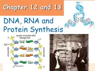
Explore the central and peripheral nervous systems, the functions of sensory and motor neurons, neuron classification, neuroglia roles, synapses, myelin function, spinal cord organization, plexuses, and reflexes. Delve into neuron organelles and the intricacies of neural circuits.

Understanding Nervous Tissue and Spinal Cord Divisions
E N D
Presentation Transcript
Chapter 12, 13 Nervous Tissue, Spinal Cord
Divisions of NS • CNS-central nervous system • Brain • Spinal Cord • PNS-peripheral nervous system- primarily nerves of body A. Spinal nerves- 31 pairs B. Cranial nerves- 12 pairs
Divisions of PNS a. Somatic- controls skeletal muscle, skin b. Autonomic-controls smooth, cardiac muscle 1. Sympathetic- fight/flight, emergencies 2. Parasympathetic-relaxation, “vegetative” reflexes
3 Basic Functions • Sensory (afferent) Receptors send impulses to CNS • Motor (efferent) CNS sends impulses to effectors (muscles or glands) 3. Integrated Functions- intelligence, creativity, personality, etc.
Organelles in a neuron • Nucleus • Granular ER(Nissl bodies) • Mitochondria • Neurofibrils(microtubules) • No centrioles -
Classification of Neurons • P365 • Anaxonic- located in brain, special sense organs • Bipolar-special sense organs • Unipolar-sensory neurons of PNS • Multipolar- most common
Neuroglia 2. Neuroglia in PNS A. Satellite Cells- similar to astrocytes B. Schwann Cells- similar to oligodenedrocytes, produce myelin sheath in PNS
Neuroglia 1.Neuroglia in CNS A. Astrocyte- function in creating blood-brain barrier, provide structure B. Oligodendocyte- produce myelin sheath C. Microglia- immune cells of CNS, similar to macrophages D. Ependymal- found in ventricles of brain, produce cerebrospinal fluid
Synapse • A specialized site where neurons communicate with one another • http://faculty.washington.edu/chudler/synapse.html
Myelin • Acts like electrical insulation • Nodes of ranvier-gaps between schwann cells on axon; allows nerve impulse to jump between nodes; leads to high conduction speeds= 100m/s • Locations- A. All motor neurons B. All spinal nerves C. 99% of brain
Unmyelinated • Slow conductions speed, .5 m/s • Located A. In autonomic nervous system
Length= 18”, width=.5” Extends from base(foramen magnum) of skull to 2nd lumbar vertebra “carrot shaped” Ends @ conus medullaris- many nerves exit and form cauda equina 2 enlargements=cervical and lumbar- where more nerves enter and leave the cord Spinal Cord- Chapter 13
# of spinal nerves-31 • Cervical- 8 • Thoracic-12 • Lumbar-6 • Sacral-5
Organization • White matter- myelinated sections on outermost parts • can be ascending- going to brain -carry sensory info -called afferent • Can be descending- coming from brain - carry motor info - called efferent
Root- where nerve enters or exits cord dorsal root=sensory/afferent ventral root= motor/efferent ** In back door out front door**
Organization • Gray matter- unmyelinated sections forming H pattern in the interior • posterior horns- contain afferent neurons • anterior horns- contain efferent neurons • “cross bar”=commissure
Plexuses • Plexus- interwoven network of nerves • Cervical plexus • Brachial plexus • Lumbar plexus • Sacral plexus
Reflexes • Rapid automatic response to a specific stimuli • Work through a reflex arc- a simple neural pathway • reception • transmission via sensory neuron • integration • transmission via motor neuron • response
Classifying Reflexes p 424 • By response A. Somatic reflex- involves skin, skeletal muscle, function in protection • Visceral reflex- involves cardiac, smooth muscle, glands, bl.v, function in homeostasis
By development A. Innate- w/drwal fr. pain, suckling, tracking objects w/ eye B. Acquired- driving, sports
3. By processing site • Spinal- patellar reflex • Cranial- sudden noise, bright light, respiration 4.Complexity of circuit • Monosynaptic- • Polysynaptic