Download
1 / 1
10 likes | 143 Vues
This study investigates the age-associated accumulation of lipofuscin granules in the myocardium of Fischer 344 x Brown Norway rats. Animals aged 6, 27, and 33 months were analyzed, including a group subjected to daily acetaminophen treatment for six months. Using confocal microscopy, we observed increased lipofuscin levels linked to aging, indicating potential oxidative damage. While chronic acetaminophen treatment appeared to diminish reactive oxygen species, it did not alter the lipofuscin accumulation in aged hearts, suggesting its role as a biomarker in cardiovascular aging.

E N D
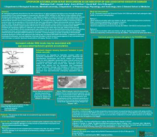
Lipofuscin granules increase with aging in the F344XBN heart. 6m 40x 6m 40x 6m 40x 27m 40x 27m 40x 27m 40x 27m 40x 33m con 40x 33m con 40x 33m con 40x 33m APAP 40x 33m APAP 40x 33m APAP 40x 33m con 40x Lipofuscin granule accumulation in 33m control Rat Myocardium (above). Lipofuscin granule Intercalated disc LIPOFUSCIN ACCUMULATION IN RAT MYOCARDIUM AS AN INDICATOR OF AGE ASSOCIATED OXIDATIVE DAMAGEMadhukar Kolli1, Anjaiah Katta1, Kevin M Rice1,2, David Neff1, Eric R Blough1,2 1 Department of Biological Sciences, Marshall university, 2 Department of Pharmacology, Physiology, and Toxicology John C Edward School of Medicine • Materials and methods: • 6-, 27-, 33-month control and 33-month Fischer 344 X BN rats which had undergone daily APAP for 6 months (30mg/kg/day) • Method 1: • Heart sections are taken with razor blade of >80 µm notice all images show ventricular fibers oriented longitudinally in the plane of section. • Incubated in Mitotracker G FM 40 nM for 15 min. • Sections analyzed by confocal microscopy (Ex-488nm / Em-522/32 nm band pass filter) • Method 2: • Heart sections are taken with razor blade of >80 µm; Sections incubated in PBS • Sections analyzed by confocal microscopy (Ex-488nm / Em-522/32 nm band pass filter) Abstract:An emerging hypothesis described as the free radical theory of aging suggests that aging occurs through the gradual accumulation of free radical damage to biomolecules (Harman, 2003). Inherent to this theory is that oxidative stress increases with advancing age. The causes of age-related increases in oxidative stress are not well understood, however it is thought to be related to declining mitochondrial function. The factor's which mediate age-associated changes in mitochondrial function are not well understood however, it is thought that age-related increases in lipofuscin may be involved since the accumulation of this molecule has been hypothesized to impair mitochondrial turnover (Brunk & Terman, 2002). Using aging Fischer 344 X Brown Norway (FBN) animals, recent studies from our laboratory suggest that aging increases reactive oxygen species (ROS) levels in these animals and that chronic acetaminophen (APAP) treatment (30 mg/kg/day) appears to diminish age related ROS levels while improving age related cardiovascular function. To test whether chronic acetaminophen may reduce ROS levels by altering mitochondria and lipofuscin bodies, we examined tissue sections obtained from the left ventricles of 6, 27, 33 month old control, and 33 month old FBN animals that had undergone APAP treatment for 6 months. Confocal imaging of cardiac preparation demonstrated increased lipofuscin content with aging and this finding was not altered with APAP intervention. Compared to traditional histological techniques, confocal mapping of lipofuscin bodies in whole mount tissues appears to be a convenient and valid technique to examine lipofuscin accumulation with aging. Harman, D. (2003). Increased cellular ROS levels may be associated with age-associated lipofuscin granule accumulation. Schematic diagram showing lipofuscin formation in post mitotic cells (left): Biomolecules are degraded by hydrolytic enzymes within the lysosomal compartment of the cell. Some macro molecules like mitochondrial cytochromes and ferritin release low molecular mass iron when degraded. Lysosomes have a low pH and are rich in reducing substances such as cysteine. Ferric iron would be reduced to the redox active ferrous form. Hydrogen peroxide, which freely diffuses into the lysosomes, may consequently undergo homolytic cleavage with formation of hydroxyl radicals which, in turn, induce peroxidative reactions and formation of lipofuscin from macromolecules undergoing degradation (Brunk 2002). Above, TEM of hamster ventricle myocardium. Dashed line surrounds lipofuscin granule. Arrow points to miochondrial cristae, dark body labeled L2 is an active lysosome stained for acid phosphatase activity. Lipofuscin granules are believed to be the end product as lysosomes break down aged mitochondria (image from Skepper 1987). Above, black arrow points to lipofuscin granules in perinuclear space; H&E preparation (image from Indiana U. Pathology) Results & Conclusions: See above, confocal imaging of cardiac preparation demonstrated increased lipofuscin content with aging and this finding was not altered with APAP intervention. In 27, 33 month optical-sections of F344 BN Rat hearts with aging the perinuclear accumulation of lipofuscin granules occurred when compared to 6 month optical-sections of rat hearts. Scale bar at top left applies to all images. Purpose:The purpose of this study is to examine the age associated changes in rat myocardium. • References: • Harman (2003). The free radical theory of aging. Antioxidant Redox Signal 5, 557-561. • Brunk, U. Terman, A. (2002). The Mitochondrial-lysosomal axis theory of aging. Eur. J. Biochem. 269, 1996-2002. • Skeeper, J.N. Navaratnam, V. (1987). Lipofuscin accumulation in the myocardium of juvenile golden hamsters: an ultrastructural study including staining for acid phosphatase. J. Anat. 150, 155-167. Specific Aim: To investigate whether chronic acetaminophen treatment may reduce the age associated oxidative damage in cardiac muscle as manifested in lipofuscin and mitochondrial morphometric changes. Hypothesis: We hypothesize that chronic acetaminophen treatment may alter the age associated oxidative damage in rat myocardium as evidenced by changes in lipofuscin accumulation. Acknowledgements: Grants for this study were supported by McNeil Pharmaceuticals to Dr Eric R Blough & Dr Ernest M Walker. Dr. M.L. Norton for maintaining the MBIC imaging facilities.