Download
1 / 30
480 likes | 1.1k Vues
Chapter 27 Molecular Fluorescence Spectroscopy

E N D
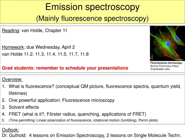
Chapter 27 Molecular Fluorescence Spectroscopy Fluorescence is a photoluminescence process in which atoms or molecules are excited by absorption of electromagnetic radiation. The excited species then relax to the ground state, giving up their excess energy as photons. One of the most attractive features of molecular fluorescence is its inherent sensitivity, which is often one to three orders of magnitude better than absorption spectroscopy.
…continued… Another advantage is the large linear concentration range of fluorescence methods, which is significantly greater than those encountered in absorption spectroscopy. Fluorescence methods are, however, much less widely applicable than absorption methods because of the relatively limited number of chemical systems that show appreciable fluorescence.
Principles of Molecular Fluorescence Molecular fluorescence is measured by exciting the sample at the absorption wavelength, also called excitation wavelength, and measuring the emission at a longer wavelength called the emission or fluorescence wavelength. Usually, fluorescence emission is measured at right angles to the incident beam so as to avoid measuring the incident radiation. The short-lived emission that occurs is called fluorescence, whereas luminescence that is much longer lasting is called phosphorescence.
Excitation Spectra and Fluorescence Spectra Because the energy differences between vibrational states is about the same for both ground and excited states, the absorption, or excitation spectrum, and the fluorescence spectrum for a compound often appear as approximate mirror images of one another with overlap occurring near the origin transition (0 vibrational level of E1 to 0 vibrational level of E0). There are many exceptions to this mirror-image rule, particularly when the excited and ground states have different molecular geometries or when different fluorescence bands originate from different parts of the molecule.
Concentration and Fluorescence Intensity The radiant power of fluorescence F is proportional to the radiant power of the excitation beam absorbed: F = K(P0 – P) where, P0 is the radiant power of the beam incident on the sample and P is the radiant power after it traverses a pathlength b of the medium. Constant K depends on the Quantum efficiency. The efficiency of fluoresced to the number absorbed.
At low concentrations where fluorescence is most often employed F = K’P0c where, c is the concentration of the fluorescent species and K’ is a new proportionality constant. F is directly proportional to analyte concentration. Thus, a plot of the fluorescent radiant power versus the concentration of the emitting species should be, and ordinarily is, linear at low concentrations. When c becomes great enough that the absorbance is larger than about 0.05, linearity is lost and F begins to reach a plateau with concentration. This effect is known as a primary absorption inner filter effect. In fact, at high concentrations, fluorescence radiant power can even begin to decrease with increasing concentration.
Molecular Phosphorescence Spectroscopy Phosphorescence is a photoluminescence phenomenon that is quite similar to fluorescence. Understanding the difference between these two phenomena requires and understanding of electron spins and the difference between a singlet state and a triplet state. Ordinary molecules exist in the ground state with their electron spins paired. A molecular electronic state in which all electron spins are paired is said to be a singlet state.
…continued… When one of a pair of electrons in a molecule is excited to a higher-energy level, a singlet or a triplet state can be produced. In the excited singlet state the spin of the promoted electron is still opposite that of the remaining electron. In the triplet state, however, the spins of the two electrons become unpaired and are thus parallel. The excited triplet state is less energetic than the corresponding excited singlet state.
Chemiluminescence Methods Chemiluminescence is produced when a chemical reaction yields an electronically excited molecule, which emits light as it returns to the ground state. Chemiluminescence reactions are encountered in a number of biological systems, where the process is often called bioluminescence. One attractive feature of chemiluminescence for analytical uses, is the very simple instrumentation. Since no external source of radiation is needed for excitation, the instrument may consist of only a reaction vessel and a photomultiplier tube. Generally, no wavelength selection device is needed because the only source of radiant is the chemical reaction.
MOLECULAR SCATTERING METHODS Several additional processes, however, can occur when radiation interacts with matter. One of the most important of these is scattering of electromagnetic radiation. Scattering can be divided into two classes: elastic scattering, in which the scattered radiation is of the same energy as the incident radiation, and inelastic scattering, in which the scattered radiation has higher or lower energy than the incident radiation. Both of these types of phenomena have useful analytical applications.
…continued… Inelastic light-scattering methods have become very important in recent years, particularly those methods based on the Raman effect. Raman spectroscopy involves inelastic scattering of radiation caused by vibrational and rotational transition. The Raman method is complementary to IR spectroscopy and can be used to obtain qualitative, structural and quantitative information about molecular species.
Chapter 28ATOMIC SPECTROSCOPY Atomic spectroscopy is used for the qualitative and quantitative determination of 70 to 80 elements. Detection limits for many of these lie in the sub-parts-per-million range. The methods can be based on absorption, emission, or fluorescence. Atomic absorption (AA) spectroscopy currently is the most widely used of these techniques.
Atomic Absorption Spectroscopy In AA spectroscopy, as in all atomic spectroscopic methods, the sample must be converted into an atomic vapor by a process known as atomization. In this process, the sample is volatilized and decomposed to produce atoms and perhaps some ions in the gas phase. Several methods are used to atomize samples. The two most important of these for AA spectroscopy are flame and furnace atomization.
Flame Atomic Absorption Spectroscopy The radiation of a line source of the element of interest, typically a hollow cathode lamp, is directed through the flame containing the atomic vapor. Solution samples are usually brought into the flame by means of a sprayer or nebulizer, which produces small sample droplets. The solvent from the droplets quickly vaporizes, and the resulting salt particles vaporize and decompose into atoms, ion and electrons.
…continued… Atoms in the sample will absorb radiation emitted by the same atom in the hollow cathode lamp and thus attenuate the power of the source. Usually a monochromator is used to separate a spectral line of the element of interest from any background radiation from the source or the flame. A photomultiplier tube is typically used to convert the radiant power from the source into a related electrical current.
Furnace Atomic Absorption Spectroscopy Furnace, or electrothermal, AA uses the same instrumental setup as flame atomizer except that a furnace atomizer is substituted for the burner. With furnace AA, discrete amounts of sample, usually microliter volumes, are deposited in the furnace. A multistep heating program is usually applied to desolvate the sample, ash, or char the organic material present and then produce the atomic vapor. Furnace AA gives a transient signal that reaches a peak in a few seconds. Furnace AA is usually one to two orders of magnitude more sensitive than flame AA.