Emission spectroscopy (Mainly fluorescence spectroscopy)
450 likes | 813 Vues
Emission spectroscopy (Mainly fluorescence spectroscopy). Reading : van Holde Chapter 11 Homework : due Wednesday, April 18 van Holde 11.2. 11.3, 11.4, 11.5, 11.6, 11.7; Midterm 2 : Friday, April 20. Overview:

Emission spectroscopy (Mainly fluorescence spectroscopy)
E N D
Presentation Transcript
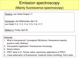
Emission spectroscopy (Mainly fluorescence spectroscopy) Reading: van Holde Chapter 11 Homework: due Wednesday, April 18 van Holde 11.2. 11.3, 11.4, 11.5, 11.6, 11.7; Midterm 2: Friday, April 20 • Overview: • What is fluorescence? (conceptual QM picture, fluorescence spectra, quantum yield, lifetimes) • One powerful application: Fluorescence microscopy • Solvent effects • FRET (what is it?, Förster radius, quenching, applications of FRET) • Linear polarization of fluorescence, rotational motion (tumbling), Perrin plots
Emission spectroscopy Conceptual Quantum mechanical picture Two (highest energy) electrons in ground state Ground state and excited states of the electrons in the outermost occupied shell of a molecule. There are different types of excited states (higher energy level states), depending on spin and angular momentum; here we will just talk about triplet and singlet states. Quick quiz: Why is quantum mechanics called quantum mechanics?
Emission spectroscopy Conceptual Quantum mechanical picture Internal conversion ~ 10-12 s Intersystem crossing ~ 10-8 s Fluorescence (wait time) ~ 10-8 s Non-radiative ~10-8 s Non-radiative ~102 to 10-11 s Phosphorcence (wait time) ~ 102 to 10-4 s Absorption ~ 10-15 s 2. singlet Absorbance, Fluorescence and Phosphorescence Different paths by which excited electrons can return to ground state – only one path results in fluorescence. Molecules will fluoresce if the emission process has a lifetime that is shorter than the conversion to the triplet state or non-radiative loss of energy. 1. singlet 1. triplet Ground state
Some Terminology • Luminescence: Process, in which susceptible molecules emit light from electronically excited states created by either a physical (for example, absorption of light), mechanical (friction), or chemical mechanism. • Photoluminescence: Generation of luminescence through excitation of a molecule by ultraviolet or visible light photons. Divided into two categories: fluorescence and phosphorescence, depending upon the electronic configuration of the excited state and the emission pathway. • Fluorescence (emission from singlet state): Some atoms and molecules absorb light at a particular wavelength, and subsequently emit light of longer wavelength after a brief interval, termed the fluorescence lifetime, t0. Fluorescent molecules are called fluorophores. • Phosphorescence (emission from triplet state): Similar to fluorescence, but with a much longer excited state lifetime.
Fluorescence spectra fluorescence Absorption h = 6.6·1034 J·s (Planck’s const.) n … frequency emission absorption In fluorescence, the return to the ground state (almost) always occurs from lowest state of excited state (0’-level).
Fluorescence spectra • Excitation:Light-induced transition, molecule goes from ground state to an excited electronic & vibrational state. • Vibrational Relaxation:Molecule loses some energy, falls to lowest vibrational state, still in excited electronic state • Emission:Molecule returns to ground electronic state while emitting a higher wavelength (=lower energy) photon
Fluorescence spectra • Excitation Spectrum: Fluorescence intensity vs. wavelength used to excite transition resembles absorbance spectrum. • Emission Spectrum: Fluorescence intensity vs. wavelength emitted for transition back to ground state: red-shifted with respect to excitation spectrum. • Quantum Yield: Ratio of energy absorbed to that emitted (more in a bit).
Fluorescence spectra Example: Fluorescein absorption and emission spectra Stokes shift White board example: Looking at the fluorescein spectrum, what is the energy difference between the ground state and the excited singlet state in fluorescein? Looking at the absorption and emission peaks: Fluorescein absorbs in the blue/purple (~ 490 nm) and emits in the green (~516 nm).
Steady State/Frequency Domain Fluorescence Instrumentation Dual monochromators, one for excitation and the other for emission. Obtain an Excitation and an Emission Spectrum.
Fluorescence Intensity t time Fluorescence decay, life-time, time-resolved fluorescence Flash sample with brief (~ns) pulse of light at lex and follow intensity vs time at lem Absorption N(0) molecules with get excited. Fluorescence intensity is proportional to number of excited molecules. • Decay of excited molecules is a first-order process, with lifetime t. • Decay can happen via three pathways: • Fluorescence with associated intrinsic lifetime to • Conversion to triplet state (phosphorescence and non-radiative decay). • Non-radiative decay.
Quantum yield When light is absorbed, only a fraction of it is emitted via fluorescence; the rest of the excited molecules decay via other processes. t is lifetime of all molecules in excited state, t0 is intrinsic lifetime (lifetime of “fluorescence state”). k, is fluorescence decay constant, A is Einstein coefficient Corollary: Fluorescence intensity is proportional to product of absorptivity (exctinction coefficient) and quantum yield.
Experimental data - fluorescence lifetime White board example: If Q is 0.5 for this fluorophore, what is the fluorescence lifetime? Gett from slope Flash lamp pulse
Factors Influencing Detected Fluorescence Intensity F(l) = Io(l)·c · e(l) · l · fF ·f(l)· d(l) F = k · c · e · fF F = k · c · S • Incident Light Intensity, Io (l) • More photons in = more photons out. Detector reads intensity, unlike absorbance. • Fraction of Light Absorbed, e(l) c l • Depends on concentration, c; path length of optical cell, l; & molar extinction coefficient, e • Quantum Yield, fF • Only emitted photons count. • Fractional Emission , f(l) • Emission at a particular wavelength • Detector Efficiency , d(l) • Wavelength-dependent instrumental factor. • Sensitivity, S • Product of extinction coefficient and quantum yield
Fluorescent amino acids • Three amino acid have intrinsic fluorescence • Fluorescence of a folded protein is mixture of fluorescence from individual aromatic residues. Most of the emissions are due to excitation of tryptophan • Tryptophan: • Highest exctinctioncoeff and highest quantum strongest fluorescence intensity. • Intensity, quantum yield, and wavelength of maximum fluorescence emission are very solvent dependent. Fluorescence spectrum shifts to shorter wavelength and intensity increases as polarity of the solvent surrounding the tryptophane residue decreases. • Tryptophan fluorescence can be quenched by neighboring protonated acidic groups such as Asp or Glu. http://dwb.unl.edu/Teacher/NSF/C08/C08Links/pps99.cryst.bbk.ac.uk/projects/gmocz/fluor.htm
Tyrosine • Like tryptophan, has strong absorption bands at 280 nm. • Tyrosine is a weaker emitter than tryptophan, but it may still contribute significantly to protein fluorescence because it usually present in larger numbers. • The fluorescence from tyrosine can be easily quenched by nearby tryptophan residues because of energy transfer effects. • Phenylalanine • Only a benzene ring and a methylene group is weakly fluorescent (product of quantum yield and molar absorbtivity maximum is low. Phenylalanine fluorescence is observed only in the absence of both tyrosine and tryptophane.
Fluorescence Excitation & Emission Spectra of Aromatic Amino Acids Absorption and Emission spectra of the aromatic amino acids in pH 7 in aqueous solutions (I. Gryczynski)
Specimen Specimen Objective lens Objective lens Collimating lens Collimating lens Dichroic mirror Dichroic mirror Hg – or Xe lamp Hg – or Xe lamp Emission filter Emission filter Excitation filter Excitation filter Camera Camera Schematics from: http://www.ifr87.cnrs-gif.fr/pbc/imagerie/outils/micros/microimg/mictout.gif Schematics from: http://www.ifr87.cnrs-gif.fr/pbc/imagerie/outils/micros/microimg/mictout.gif Powerful application of fluorescence: Fluorescence microscopy(shown here: epi-fluorescence illumination) Note: Photobleaching: Fluorophores will “die” after a while of intense illumination.
Fluorescence microscopy Normal African Green Monkey Kidney Fibroblast Cells (CV-1) (from Olympus web page: http://www.olympusmicro.com). Immunofluorescently labeled with primary anti-tubulin mouse monoclonal antibodies followed by goat anti-mouse Fab fragments conjugated to Rhodamine Red-X. In addition, the specimen was stained with DAPI (targeting DNA in the nucleus).
Fluorescence microscopy • Advantages: • Can label selected features of a sample, eg. Nucleus, DNA, microtubules, specific proteins • Can observe how those molecule behave over time. • Can see (though not resolve) features on nanometer level, even single molecules.
Solvent effects Solvents can affect the fluorescence emission spectrum hugely. Emission wavelength can be shifted and quantum yield can change. There are specific and general solvent effects. Specific solvent effects: A chemical reaction of the excited state with the solvent. Example: Hydrogen-bonds, acid-base interactions, charge transfer. Changing Fluorescence can be used to detect solvent interactions. Specific solvent interaction: 2-anilinonaphthalene fluorescence was changed to high wavelength by replacing cyclohexan with ethanol. Ethanol forms hydrogen bond.
Solvent effects • General solvent effects: Fluorescence depends on polarizability of solvent • Increasing dielectric constant of solvent shifts usually fluorescence to higher wavelength. • Polar interactions lower energy of excited state (stabilize excited state). Lower DE: DE = hc/l Emission at longer l Polarization Red-Shift 0rg H20 Putting a fluorophore from cyclohexan (low dielectric constant) into water (high dielectric constant), shifts fluorescence to higher wavelengths.
Quantum yield depends very much on environment Changing quantum yield upon binding Increased quantum yield upon binding Application: Staining of DNA in gels. Fluorophores with good DNA binding affinities (often intercalation), extremely large fluorescence enhancements upon binding nucleic acids (some >1000-fold), and negligible fluorescence for the free dyes. Qrel = 1.00 Qrel = 0.46 Qrel = 0.23 SYBR stained dsDNA gel. Excite with UV, emits in visible. (DNA/SYBR Green I complex: Q~0.8; ~300-fold increase over free dye)
Fluorescence resonance energy transfer (FRET) • When two fluorophores are close together it is possible that one of them absorbs the light (donor), then transfers the energy to the neighboring fluorophore (acceptor), which then emits the light. • The two conditions for this to happen are: • Transition dipole interaction between the two fluorophores (i.e., they need to be close together and aligned). • Significant overlap of the emission spectrum of the donorwith the absorption spectrum of the acceptor. Example: Fluorescein (donor) and Alexa-546 (acceptor):
The absorption and emisson spectra of some fluorophores Good FRET pair
Fluorescence resonance energy transfer (FRET) Efficiency of transfer: Basically, FRET is a great method to determine the distance between two fluorophores (molecules) in the range of ~1-10 nm. r … D-A distance R0 Förster radius Close together FRET signal Far apart (further than Förster radius) no FRET signal • Clever example: Molecular Beacons (also: more later on quenching) • used to detect presence of a certain DNA sequence in solution or cells (show on white board).
Fluorescence energy resonance transfer (FRET)Donor-acceptor pairs • White board example: • What is the FRET efficiency for a fluorescein-rhodamine pair, that is 2.25 nm apart? • If excited with 488 nm light, what is the main emitted wavelength?
FRET Application: Coexisting conformations of fibronectin in cell culture imaged using fluorescence resonance energy transfer, Baneyx et al., PNAS 98:14464, 2001 D A Extended conformation: Donor and acceptor are far apart: no FRET (donor emits) Folded conformation: Donor and acceptor are close FRET (acceptor emits)
FRET Detects Compact and Extended Fibronectin Conformations in Cultured Cells
Quenching Some molecules are quenchers they suppress fluorescence when they are close to a fluorophore Fluorophore = fluorescein Quencher = dabcyl
Quenched no light Not quenched light Molecular Beacons & Fluorescence Quenching
Linear polarization of fluorescence • Light to excite fluorophore is now linearly polarized • Emitted fluorescent light will be depolarized Absorption is best for those molecules whose transition dipole is parallel to plane of polarization. • (De-)Polarization of emitted light depends on: • Orientation of emitting transition dipole relative to absorbing transition dipole • Amount of molecular rotation during fluorescent lifetime! • Depolarization of emitted light
Linear polarization of fluorescence Depolarization is described in terms of: • Assume molecules don’t rotate while being excited • depolarization due only to random orientation of molecules with respect to incoming light, q, and angle g: Anisotropy for fluorescence of rhodamine as a function of l of exciting light If there is no molecular rotation, anisotropy will vary between 2/5 (absorbing and emitting trans. dipoles are parallel) and-1/5 (dipoles are perpendicular).
Linear polarization of fluorescence Fluor. anisotropy r r time • Now assume molecules tumble (rotate) before emitting. • depolarization due rotation of molecules. Two extremes: i) molecules don’t rotate before emission r = r0 ii) molecules randomly orient before emitting: r = 0 Time-resolved fluorescence provides a convenient way to measure rotational motion of biological molecules. • … correlation time information about size &shape of molecule • large slow tumbling large molecular weight
Example: rotational correlation time for BSA Mol Wt = 66,500 grams/mole Drot = 8.3E6 sec-1 How long for BSA to “tumble”? Rotational Correlation Time: r= V h / k T = (8.1E-20)*(0.01) / [1.38E-16 * 293] r= 2.0E-8 sec = 20 nsec BSA rotates in ~ 10 - 20 nanonseconds Fluorescent lifetime ~ 5 nanoseconds
Linear polarization of fluorescence Large r slow rotation large molecule Small r faster rotation compact molecule
Perrin plots Instead of pulse illumination, use continuous illumination to measure anisotropy will get average anisotropy ravg. HW 11.6 • … lifetime h … viscosity T … temperature V … volume of molecule
Applications of fluorescence to proteins • Fluorescence microscopy • Analytical detection of presence of proteins • Monitor changes in quantum yield as indication of changing environment (binding, unfolding, etc.) • Effects of energy transfer (FRET). Determine distance of fluorescent groups from each other in 1-10 nm range. • Changes in fluorescence polarization to determine shape and size of molecules (tumbling depends on shape and size) • Monitor (change) in fluorescence parameters to determine stoichiometry, presence of intermediates, binding constants, etc.
Application of fluorescence to DNA • Staining of oligonucleotides in gels • Monitoring the unwinding of double-stranded DNA helicase • Monitoring DNA melting Also: there are tons of reactive fluorophores that can be used to label proteins (Cysteines, primary amines, etc) and DNA. See: Molecular Probes, Inc. http://probes.invitrogen.com/