Download
1 / 13
140 likes | 435 Vues
The Lung and Respiration 2/24. What are the major anatomical features of the respiratory system. How is air cleaned prior to gas exchange? How is air distributed into smaller and smaller airways? What special structures in a trachea are related to the function of a trachea?

E N D
The Lung and Respiration 2/24 What are the major anatomical features of the respiratory system. • How is air cleaned prior to gas exchange? • How is air distributed into smaller and smaller airways? • What special structures in a trachea are related to the function of a trachea? • How do cilia clean dirt and other materials from the lung? • How does alveolar structure promote gas exchange?
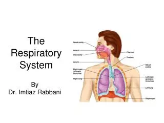
The Respiratory System provides for gas exchange between the blood and air. • “Respiration” has three different connotations: • 1) Lung Ventilation: • 2) Lung Gas Exchange: • 3) Tissue: oxygencarbon dioxide and energy • Upper Respiratory Tract: Head and Neck • Filtering out harmful particles in “air” • Humidifying “air” • Delivering “air” to the lung • Lower Respiratory Tract: Thorax • Gas Exchange • Prevention of infection • Clean alveolar surface • Ventilatory Mechanics: Skeletal muscle of diaphragm and intercostal muscles between ribs. Also: abdomenal muscles • Most people only use energy to “inhale”! (Exhale for free)
What organs facilitate the movement of air to the lung? The importance of mucus and the mucus membrane: CRITICAL! • Air Entry: • Nasal Cavity and Conchae- • Sinus Cavities- • Oral Cavity: poor airflow efficiency/low resistance • Pharynx- • Epiglottis and Glottis- • Trachea- • Bronchial Tree- • Alveolus- • Each lung and its lobes sit in a pleural cavity under a slight vacuum • Esophogus is often mistaken for an airway during anaesthesia, this can be fatal!
Overview of the primary airway structures. Each lung (Rt/Lt) is surrounded by pleural fluids and sits in a pleural cavity that is under a slight vacuum. HEART V.I.P: Pleural Cavity under vacuum
Air is distributed into lower respiratory air passages called bronchi and bronchioles that ultimately lead to dead-end alveoli. “The Bronchial Tree”: moves air from large to narrow branching airways • Bronchi with cartilage for support and smooth muscle: • Primary BSecondary BTertiary BBronchioles • Have cilia for cleaning purposes • Nourished by bronchiolar artery • Bronchioles are 1mm diameter airways with smooth muscle for changing diameter to needs BronchioleTerminalBronchioleRespiratoryBronchiole Alveoli(Gas Exchange) Respiratory Bronchiole and alveoli have no cilia or smooth muscle! The smooth muscle lining Bronchiole can constrict Asthma • Alveoli: clusters of dead end pouches for gas exchange! • Alveolar macrophages: cleaning • Type II alveolar cells: surfactant • Type I alveolar cells (simple squamous epithelial): gas exchange
Bronchiole Terminal Bronchiole Respiratory Bronchiole Alveolar Sacs
Amazing Statistics about the lung: • A fully inflated lung can contain about 5-6liters of air. • The surface area of the 150 million tiny alveoli in each lung is about 75 meters2 • The surface area of the capillaries that sit underneath the alveoli is about 60 meters2 for a single lung. • Surprisingly the volume of blood in these alveolar capillaries at rest is only about 70 ml and perhaps 200 ml when exercising rigorously. • Clearly the blood must move through these capillaries very rapidly and gas exchange must ALSO be very rapid (transit time across the alveoli is 0.75 seconds or less).
The shape of the lung describes why we sometimes have respiratory difficulties. • Each lung sits in a pleural cavity, you have two pleural cavities in your thorax. • Why do right and left lung lobes have different sizes? • Importance of having 2 lobes sealed separately? • Pneumothorax: if pleural cavity loses its vacuum, the lung collapses and gas exchange to blood stops! • Implications:Cancer? Infection? GunShot (deer/human)? • Why do aspirated objects/obstructions affect the right side more often than the left? A story about kids eating marbles! • If you have to have a lung removed, why does left lung removal affect you less than right lung removal?
The respiratory system is rich in elastin and cartilage for two very different reasons. • Why is elastin so important? • It stretches and stretches back! • Located in lung tissue and thoracic wall! • Inhalation: Active Exhalation: Passive • Emphysema/cigarette smoke/ alpha-1-antitrypsin/elastin loses • Barrel Chest: sign of loss of elastin • Why is cartilage so important? • Hyalin Rings: trachea and bronchi of lung • Cartilagenous rings prop airways open and keep airways from collapsing when you inhale • Smooth muscle in airways lets you control tune diameter/airflow • Cartilaginous hyalin rings help prevent airway collapse when you INSPIRE! Why? • Elastin causes the lung to collapse passively (exhale)! WHY?
Asthma: trachealis and other airway smooth muscle cells constrict in response to irritants, this increases airway resistance. Q α r4 V.I.P Changes in resistance are most pronounced when the bronchioles constrict. Asthma can also be “protective” sometimes….why?
Cilia are tiny “hair-like” structures that carry mucus, bacteria, pollen, dust and other potentially harmful material (caught in mucus) up and out to the glottis. However, cilia do not extend into the alveolus/terminal bronchiole and can be destroyed by smoke! Why do you sometimes have to “cough”? Why do smokers cough more often?
Lungs move and circulate air to maximize alveolar gas exchange. Unfortunately we are not efficient at doing this. • Primary, Secondary and Tertiary Bronchi -Cilia, SMC, non-respiratory and brachial artery -Acute effects of asthma found here! • Terminal Bronchioles and Respiratory Bronchioles • ------Scant-gas exchange/ “no” or “few” cilia /No or few SMCs • Until this point the air sits in what can be called a “dead space” (called dead because significant gas exchange can’t occur here). These dead spaces should never collapse. • You CAN inhale air and get no gas exchange to the blood because of collapsed bronchioles due to asthma!
Terminal and respiratory bronchioles have very short lengths that terminate in the blind alveolar sacs. Each sac is richly supplied with a capillary bed for gas transfer to/from blood.