Microtubules (MTs)
Microtubules (MTs) . E. D. Salmon Biology tsalmon@email.unc.edu Reading: Lodish et al., Molecular Cell Biology; Alberts et al., Molecular Biology of the Cell. Some Microtubule Functions. Provide structure, acting as an internal skeleton


Microtubules (MTs)
E N D
Presentation Transcript
Microtubules (MTs) E. D. Salmon Biology tsalmon@email.unc.edu Reading: Lodish et al., Molecular Cell Biology; Alberts et al., Molecular Biology of the Cell
Some Microtubule Functions • Provide structure, acting as an internal skeleton • Act as a polarized tracks for microtubule motor (cytoplasmic kinesins and dyneins) driven movements within cells • With axonemal dynein produce motility of cilia and flagella • With microtubule motors segregate chromosomes in mitosis • Give cell polarity and produce polarized organization of organelles • Regulate activity of actin cytoskeleton in cell motility and cell division
The Tubulin Dimer • Each tubulin is about 50 kD and structurally very similar to each other and to FtsZ in bacteria • Microtubules are composed of a and b tubulins b • Each tubulin binds GTP: the a GTP is non-exchangeable and the dimer is very stable, Kd = 10-10; the b GTP is exchangable in the dimer • The intracellular tubulin concentration is about 20 mM; about half in microtubules and half as dimers in the cytosol a
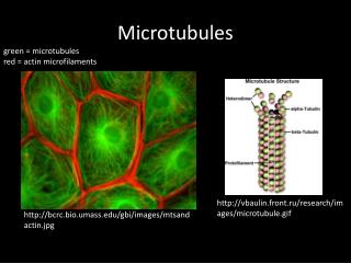
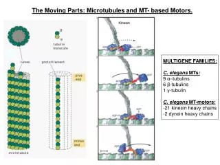
Microtubules are Polarized Polymers of Tubulin + • a/b heterodimers bind head-to-tail along protofilaments • 13 protofilaments form a hollow tube-the microtubule: 25 nm OD, 14 nm ID • Microtubules are polar-they have a plus and a minus end • *1625 dimers/mm length • *A mammalian tissue cell (6 pL) has 16,500 mm total microtubule length; a frog egg (1 mL) has 2,500 m -
Other Microtubule Geometries Axomeme (Cilia and Flagella) Cytoplasmic: (10-16 pf) 13 pf is typical Centriole or Basal Body
Microtubule Organization in Interphase and Mitosis Centrosome 5 mm 25 mm
Polarized Microtubule Organization in vivo Centrosome + + + + + + + + + Mitosis Interphase
Microtubules are Abundant in Neurons:Brain is the best source of tubulin - +
Microtubule Assembly Occurs By End- Dependent Association-Dissociation Reactions a b • In vitro, microtubules that self-assemble from pure tubulin • can grow and shorten at both ends • The plus end grows faster and is more dynamic than • the minus end
Microtubule Self-Assembly From Pure Tubulin In Vitro: • 2 mg/ml tubulin in 1 M PIPES, 1mMEGTA, 1mM MgCl2, 1mM GTP pH = 6.8 and 37C (can “seed” MTs using preformed cross-linked MT fragments for example: Cc [tub] in polymer
Microtubule Ends Exhibit Dynamic Instability, Not Simple Equilibrium Assembly + End
b b At an end: dL/dt ~ [fg(kag(S-Ccg)) –fskds]
Microtubule Ends Change Conformation Between Growth and Shortening Phases
Recording Microtubule Dynamics in Living Cells Or, express Tubulin-GFP
Hamamatsu Orca ER CCD Camera • Low readout noise (~8 electrons) • High Quantum Efficiency • Broad spectral response • Fast readout: ~14MHz
Yokogawa Scanning Head Nikon TE300 inverted microscope Filter Wheel Orca ER CCD Focus Controller 80 mW Argon-Krypton Laser Input (fiber optic)
Microtubule Polarity and DynamicsDynamic Instability In vitro mitotic cell extract + centrosome Living interphase animal cell; cell edge Fluorescent Speckle Microscopy MTs green; actin red (Salmon and Waterman)
tubulin Ring Complexes Nucleate Plus-End Growth in Cells; Self-Assembly and Minus-End Growth Is Rare + gTubulin Ring Complex g g gTuRC~12-14 gtubulins, Xgrips72,109,110,133,195 g - g g g g g g g g Zheng et al. 2000 Nat. Cell Biol. 2:358
Centrosome is A Microtubule Organizing Center (MTOC) MTOC’s control where microtubules are formed Centrosomes contain peri-centrosomal nucleation complexes surrounding pair of centrioles Centrioles within centrosomes become basal bodies, which are nucleation centers for cilia and flagella
Centrosome is a Microtubule Organizing Center (MTOC) Centrosomes contain: Peri-centrosomal: gTuRC nucleation complexes bound to pericentrin and centrin fibrous material plus many kinases
Microtubule Associated Proteins (MAPs) Control Microtubule Assembly and Many Stabilizing MAPs Bind to Outer Surface of Protofilaments
Catastrophe Factors and Rescue Factors: Many Concentrate at MT Plus Ends • Catastrophe: Op18, Stathmin; End-binding protein • Kin I (e.g.XKCM1, hMCAK); Kip3 • Rescue: Stabilizing Microtubule Associated Proteins (MAPs) • -XMAP215 (hTog, S.c. Stu2 ): antagonizes KinI • -Brain Maps (MAP2, Tau) • -Growing End Binding proteins: EB1,CLIP170 • *Note: Activity of all these factors regulated in the cell cycle • by phosphorylation or during cell motility • *Note: In budding yeast, microtubule motors can effect plus • end dynamics: destabilize-Dyn1, Kar3, Kip3 • stabilize-Kip2
MT Assembly Dynamics is Regulated in the Cell Cycle (Cyclin B/Cdk1 kinase) (Inactive) (Active) Cat. Rescue
Proteins Which Transiently Bind Growing Ends • EB1: Links to APC which can bind Beta catenin at cortex; EB1 binds Ncd kinesin motor • Bim1 in yeast binds Kar 9 at cortex • Clip 170: Links to dynein/dynactin, CLASPS • Dynein/dynactin-links to cortex (binds beta-catenin • Ncd: minus kinesin • MCAK: KinI depolymerase • Review: Vaughan KT, 2004, Surfing, regulating and capturing: are all microtubule-tip-tracking proteins created equal? Trends Cell Biol. 2004 14:491-6.
Alexa-488-EB1 Bound to the Growing Ends(10 mm/min)of Microtubulesin Early PrometaphaseSpindle in Xenopus EggExtracts Jennifer Tirnauer
Microtubule Drugs Bind tubulin dimer and blocks assembly: Colchicine, nocodazole, vinblastine, podophilotoxin, vincristine Binds dimer in microtubule lattice and stabilizes microtubules: Taxol