Thrombophlebitis and Occlusive Arterial Disease
490 likes | 2.21k Vues
Thrombophlebitis and Occlusive Arterial Disease. October 6 th , 2005 George Filiadis D.O. . Thrombophlebitis. Formation of a venous clot depends on the presence of of at least of one of Virchow’s triad factors -venous stasis -injury to vessel wall -hypercoagulable state.

Thrombophlebitis and Occlusive Arterial Disease
E N D
Presentation Transcript
Thrombophlebitis and Occlusive Arterial Disease October 6th, 2005 George Filiadis D.O.
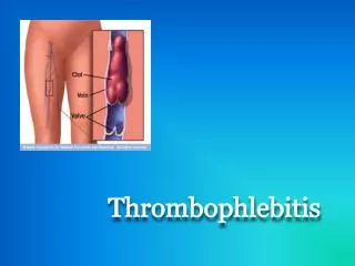
Thrombophlebitis • Formation of a venous clot depends on the presence of of at least of one of Virchow’s triad factors -venous stasis -injury to vessel wall -hypercoagulable state
Trauma, travel Hypercoagulable, hormone replacement Recreational drugs(IV drugs) Old (age >60y) Malignancy Birth control, blood group A Obesity, obstetrics Surgery, smoking Immobilization Sickness Clinical risk factors for deep vein thrombosis
Pathophysiology • Most common cause of hereditary hemophilia is factor V Leiden • See Table 59.2 for other hypercoagulable states • Thrombi usually form at the venous cusps of deep veins where altered or static blood flow causes clot formation • Alternatively, clots form from intimal defects • Clots are composed from fibrin, red cells and platelets and cause partial/complete obstruction of vein
Pathophysiology • Postphlebitic syndrome (PPS) may develop after the resolution of a DVT • PPS is due valvular incompetence, persistent outflow obstruction and abnormal microcirculation.
Superficial Thrombophlebitis • Thrombosis can occur in any superficial vein primarily the saphenous vein and its tributaries • Local pain, redness, and tenderness are characteristic findings. • Mild cases can be treated with warm compresses, analgesia and elastic supports • Severe cases can be debilitating and should be managed by bed rest, elevation of extremity, support stockings, and analgesia. • Antibiotics and anticoagulants are useful in septic thrombophlebitis
Superficial Thrombophlebitis • Incidence of DVT from extension of a superficial clot is 3%. • Most clots in great saphenous vein will extend into a deep vein system in a week or so thus a follow-up US is guaranteed • Definite treatment is ligation and excision of affected vein.
Deep Vein Thrombosis • Clinical exam is unreliable for detection or exclusion of a DVT • Pain, redness, swelling, and warmth are present in less than half the patients with confirmed DVT. • Pain in calf with dorsiflexion of ankle with the leg straight (Homan’s sign) is unreliable • See table 59.3 for predictors of deep vein thrombosis
Deep Vein Thrombosis • Symptomatic DVT will be in popliteal or more proximal veins more than 80% • Nonextending calf DVT rarely cause PE • Uncommon presentations of DVT include phlegmasia cerulea dollens and phlegmasia alba dollens • In phlegmasia cerulea dollens, patients present with extensive swollen and cyanotic leg due to massive ileofemoral thrombosis which can lead to venous gangrene.
Deep Vein Thrombosis • In phlegmasia alba dolens, the leg is white due to arterial spasm secondary to massive iliofemoral thrombosis, often mistaken for arterial occlusion. • PPS can be difficult to differentiate from recurrent DVT due to pain, swelling and ulceration of the skin. • Up to to one third of the patients with DVT can develop PPS.
Deep Vein Thrombosis-Diagnosis • All patients with any signs or symptoms suggestive DVT should undergo an objective diagnostic evaluation • Venography was the historical “gold standard” for detection of DVT with 100% sensitivity and specificity but it is invasive and can cause contrast-related reactions, phlebitis and DVT (3%).
Deep Vein Thrombosis-Diagnosis • Choice of test to identify DVT is ultrasound • Ultrasound has 97% and 94% sensitivity and specificity respectively for detecting proximal DVT • Ultrasound is less sensitive for pelvic DVT and has sensitivity of 73% for a calf DVT. • Impedance plethysmography is portable and inexpensive but less sensitive than US • IP measures changes in electrical resistance in response to changes in calf volume due to obstruction
Deep vein thrombosis-Diagnosis • Radioisotopes have been used to diagnose DVT but are not particularly useful in ED • MRI is being used with increased frequency and can detect a filling defect in entire extremity (including calf and pelvic veins) • D-Dimer fragments which are degradation products of fibrin can be used to as an indicator for the presence or absence of DVT or PE. • The ELISA based D-Dimer has sensitivity 97 % and specificity 35%
Deep Vein Thrombosis-Diagnosis • When D-Dimer is less than 500ng/ml, the likelihood of DVT is less than 1%. • The latex agglutination assay D-Dimer is less sensitive than the ELISA essay. • Sepsis, surgery, trauma, hemorrhage, pregnancy, cardiovascular diseases, collagen vascular disease, liver disease, cancer are associated with elevated d-dimer value.
Treatment • Bed rest, leg elevation and elastic stockings are of unproven benefit in the management of DVT. • Aggressive anticoagulation will prevent extension of the clot. • Early ambulation after adequate anticoagulation is a safe approach • Primary objective of treating DVT is the prevention of pulmonary embolus
Treatment • Patients with negative ultrasound can safely have a repeat ultrasound in a week without anticoagulation • Risk of PE in these patients is near 0% and risk of forming a DVT is 1%. • Anticoagulation is recommended for patients with calf DVT who had PE/DVT, immobile, have hypercoagulable state
Treatment • Patients with proximal DVT require anticoagulation • Preferred treatment is LMWH over UFH because of the ease of administration, more predictable anticoagulant effect, lack of need to monitor the anticoagulation effect, lower incidence of major bleeding and HIT • LMWH has a preferentially inhibitory effect on factor Xa.
Treatment • Because of LMWH is cleared by the kidneys, it should be avoided in outpatients with Cr >2.03 • One need not to wait for the creatinine result before initiating LMWH therapy. • The ability to discharge patients from the ED after initial dose of LMWH is cost-effective, safe, practical and acceptable practice as long as there is a secured 24 hr follow up with PCP.
Treatment • Indications for admission include inability to ambulate, poor social support, unreliable follow-up, difficulty with education with drug administration, need for lysis or invasive therapy, and an alternative serious diagnosis under investigation or that requires treatment(arterial ischemia, cellulitis, pelvic mass)
Treatment • If LMWH is contraindicated, use UFH as 80 units/kg bolus and then 18 units/kg/hr • Serious bleeding from LMWH cannot be completely reversed with protamine which has been associated with hypotension and anaphylactoid reactions. • If a patient has contraindication to heparin like in pt with HIT, you can use a thrombin inhibitor like lepirudin
Treatment • In pregnant pt who cannot have heparin, danaproid should be used. • It is acceptable to start coumadin and LMWH simultaneously. • Warfarin is contraindicated in pregnancy, active bleeding, recent major surgery (thoracoabdominal, nervous system, spine, eye) • LMWH does not interfere with the work up of a possible hypercoagulable state compared with UFH.
Treatment • Initial hematological testing at follow-up includes factor V leiden, prothrombin molecular tests, screening for antiphospholipid anticoagulants and a fasting homocysteine level. • Upon completion of the anticoagulation , further testing includes antithrombin III, protein C, protein S, and factor VIII level
Treatment • Thrombolysis for DVT is indicated for extensive iliofemoral thrombosis and upper extremity DVT in patients with low risk for bleeding. • IVC filter is indicated for when anticoagulation therapy is contraindicated, there is embolization of DVT after 1-2 weeks of anticoagulation
Treatment • Thrombectomy is only indicated with ischemic leg secondary to a massive venous clot like in phlegmasia cerulea dolens. • In ED , pt adequately anticoagulated who present with new thrombus or propagation should receive LMWH • If the fail LMWH or there is a free-floating thrombus an IVC should emergently inserted.
Pelvic Vein Thrombosis • Usually it’s an extension of a clot from the femoral vein. • An isolated pelvic vein thrombosis is rare and can be a complication in the postpartum period, after pelvic surgery or trauma. • Septic pelvic vein thrombophlebitis is a life-threatening condition after post-partum endometritis and is usually diagnosed with CT or MRI.
Axillary and Subclavian Vein thrombosis • 2-4% of DVTs occur in axillary or subclavian vein • Risks include recent central venous catheters or pacemakers, IV drug use, malignancy, hypercoagulable states and excessive or unusual exercise, chronic compression(cervical rib, scalene or web) • PE occurs in 5-10% of cases involving axillary or subclavian DVT • Treatment includes anticoagulation alone or preceded by thrombolysis.
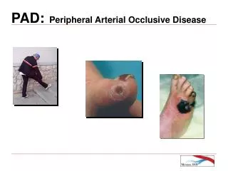
OCCLUSIVE ARTERIAL DISEASE • Acute limb ischemia secondary to thrombosis or embolus is true emergency. • Mortality is 25% and risk of amputation is 20%. • 11-27%of elderly have peripheral arterial disease • Smoking, diabetes, hyperlipidemia, hypertension and homocysteinemia are significant risk factors • At least half of the patients with coronary or cerebrovascular disease have PVD
Pathophysiology • Acute limb ischemia leads to cell death and irreversible tissue damage. • After prolonged arterial obstruction, reperfusion may not be fully attainable due to distal edema and thrombi forming in the microcirculation. • Peripheral nerves and skeletal muscle are very sensitive to ischemia and irreversible damage can occur within 6 h of anoxia • Non-embolic ischemia is due to atherosclerosis of the vessels
Pathophysiology • Progression of ischemic injury can occur through several mechanisms: i)propagation of clot to include collateral vessels ii)ischemia-related distal edema leading to high compartment pressures iii)fragmentation of clot in the microcirculation iv)edema of the microvasculature cells
Etiology • Thrombotic occlusion is significantly more common cause of acute limb ischemia than is embolism. • Emboli originate from the heart in 80-90 % with atrial fibrillation being the cause in two thirds of all peripheral emboli. • Mural thrombus in the ventricle after recent myocardial infarction is the second most common cause.
Etiology • Other causes of emboli include atrial myxomas, vegetations from valve leaflets, and parts of prosthetic devices such as mechanical valves. • Noncardiac causes include thrombi from aneurysms and atheromatous plaques. • Iatrogenic embolization can happen during angiograhic procedures of the aorta and larger vessels
Etiology • Thrombosis unrelated to atherosclerotic disease can occur at an area of vessel injury during invasive studies. • Peripheral arterial supply can be obstructed by vasospastic or inflammatory conditions like Raynaud disease and Thromboangitiis obliterans (young smokers) • Limb ischemia can also seen with central causes like thoracic aortic dissection and Takayasu arteritis.
Etiology • Low cardiac output states like cardiogenic or hypovolemic shock may also present with limb ischemia • Cardiac tamponade, ischemic cardiomyopathy, valvular heart disease can impair left ventricle function and lead to leg ischemia in patients with existing peripheral vascular disease.
Clinical features • 6 Ps:pain, pallor, polar (for cold), pulselessness, paresthesias, and paralysis. • Despite the belief that the limb salvage is possible within 4-6h, tissue loss can occur with significantly shorter occlusion times. • Chronic peripheral arterial insufficiency is characterized by intermittent claudication with activity that is relieved at rest. • Shiny, hyperpigmented skin with hair loss and ulceration, thickenend nails, poor pulses are hallmarks of chronic disease
Diagnosis • Clinical evaluation is the most useful diagnostic tool. • Capillary refill is not reliable alone • A hand –held Doppler can detect the presence or absence of a pulse. • If a pulse is detected, then the ankle-brachial index (ABI) and segmental leg pressures should be checked • An ABI<0.5 indicates acute arterial obstruction • If time permits, do a duplex ultrasound
Treatment • Goals of therapy include restoration of blood flow, preservation of limb and life, and prevention of recurrent thrombosis • Current practice includes UFH to prevent clot extension, venous thrombosis, the appearance of thrombi distal to the obstruction, and reocclusion. • Fluid resuscitation and treatment of heart failure and dysrhythmias are sometimes necessary to improve limb perfusion. • Definite treatment includes surgery or thrombolysis
Upper Extremity Ischemia • Upper extremity arterial occlusion is less common. • There is a well-developed collateral circulation around the shoulder and elbow, thus arterial occlusion is better tolerated. • Usual causes are vasospasm, arteritis, trauma, hypercoagulable state, plaque rupture, thoracic outlet syndrome, aneurysms. • Treatment includes heparinization and surgical thrombectomy.
Aneurysms of the extremity • Incidence of aneurysms in lower extremities appears to be increasing due to the aging population. • Femoral and popliteal aneurysms are the most common. • Symptoms include local pain, limb edema, and ischemic complications • For femoral aneurysms (majority are false), US, CT or MRI can confirm the diagnosis
Aneurysms of the extremity In patients with popliteal aneurysm there is 37% chance of abdominal aortic aneurysm and 50 % chance of coexisting popliteal aneurysm on the contralateral leg. Subclavian artery aneurysms can produce central neurologic findings or upper extremity iscemia and are due to atherosclerosis, trauma, thoracic outlet obstruction, syphilis or cystic medial necrosis.
Questions • 1.Regarding superficial thrombophlebitis, all of the following are correct except: • A)treatment includes bedrest, elevation, support stockings, and analgesia. • B)antibiotics and anticoagulants are of no proven benefit. • C)incidence of DVT from superficial thrombus is 30% • D)all of the IV drug users with superficial DVT should receive antibiotics
Questions • 2.T/F Calf DVT usually extend proximally and are a common case of PE • 3.T/F Occlusive arterial disease is usually due to a thrombosis event rather than embolic • 4. T/F Pt with political aneurysm frequently have abdominal aortic aneurysm and contra lateral political aneurysm.
Questions • 5.Which of the following statements is true • A)Diagnosis of a DVT can be based on clinical exam • B)Test of choice for diagnosis of DVT is ultrasound. • C)Venography is the gold standard diagnostic modality given its low complications rate • D)Ultrasound has higher sensitivity for detecting calf DVT than proximal DVT • Answers: 1)c, 2)F, 3)T, 4)T, 5)B