Peripheral Artery Occlusive Disease
440 likes | 646 Vues
Learn about risk factors, symptoms, diagnosis, prognosis, and treatment of Peripheral Artery Occlusive Disease (PAOD) from Dr. Mehdi Hadadzadeh, a cardiovascular surgeon. Find out about its prevalence and associated cardiovascular risks.

Peripheral Artery Occlusive Disease
E N D
Presentation Transcript
Peripheral Artery Occlusive Disease Dr.mehdi hadadzadeh Cardiovascular surgeon
Prevalence • Approximately 1 million Americans become symptomatic Q year • Approximately 5% of men and 2.5% of women complain of intermittent claudication by history • If asymptomatic disease is included (as determined by ABI) 13% of women and 16% of men have peripheral vascular disease • Of these only 1% have critical limb ischemia
Risk Factors • Age • Male gender (over age 70 risk equalizes) • DM (tend to have more distal and diffuse disease; 7 fold increase risk of amputation) • Tobacco (risk even stronger than for CAD; with smokers experiencing IC up to 10 yrs earlier) • HTN • Hyperlipidemia
Prognosis • Over 5-10 yrs 70% of pt’s have no change or improve • 20-30% worsen • 10% require intervention • 1% require amputation • In patients with IC the majority of morbidity and mortality comes from increased risk of CAD/CVD
Associated Risks (CAD/CVD) • Estimated that of those with lower extremity arterial disease at least 10% also have CVD and 28% have CAD • Of patient with LE arterial disease 75% will die of a coronary or cerebrovascular event
History • Quality (aching, numbness, weakness, fatigue) • Location (calf, buttock, or thigh) • Severity of pain and functional limitations • Typically induced by walking and relieved by rest • True claudication typically resolves in <10 minutes after stopping activity • Nocturnal pain and pain at rest are indications of more severe disease • Risk Factors
Physical Exam • Condition of skin and appendages • Pulses • Check for bruits • Pallor during leg elevation • Time for color return after leg restored to dependent position • ABI
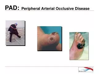
Symptoms • Intermittent claudication • Rest pain • Erectile dysfunction • Sensorimotor impairment • Tissue loss Signs • Muscular atrophy • Decrease hair growth • Thick toenails • Tissue necrosis ulcers infection • Absent pulses • Bruits
Clinical Presentations of PAD ~15% Classic (typical) claudication 50% Asymptomatic ~33% Atypical leg pain(functionally limited) 1%–2% Critical limb ischemia
Ankle Brachial Index (ABI) • ABI <0.9 is 99% sensitive and 99% specific for angiographically diagnosed PAD • Supine position • Check systolic BP in upper extremities (using Doppler) – use highest value • Systolic BP in lower extremities – use highest value • Divide ankle SBP by brachial SBP • May be falsely elevated in calcified vessels (DM)
ABI • Normal = >0.90 • 0.70 – 0.89 = mild disease • 0.50 – 0.69 = moderate disease • <0.50 = severe disease (rest pain/tissue loss) • If strongly suspect IC but WNL, can repeat following exercise (leg pressures only)
Other Noninvasive Testing • Segmental Pressure Measurements • Pulse Volume Recordings • Duplex Scanning • MRA
Segmental Pressure Measurements • Measures SBP at multiple levels (upper and lower thigh, upper calf, ankle) • Pressure reductions between levels help to localize occlusion • Normally pressures increase as move further down the leg (>20mmHg gradient abnl) • Limited with calcified artery walls (ie: diabetics)
Pulse Volume Recordings • Pneumatic cuffs placed similarly to SPM with pulse volume recorders • Calibrated air plethysmographic wave form recording system • Instead of SBP, measure volume of blood entering the arterial segment during systole • Generates a waveform which normally has rapid systolic peak and dicrotic notch • Not limited by calcifications of vessel walls
SPM and PVR • Useful in measuring general local and severity of obstruction • Allow for objective monitoring of patient’s change over time through serial exams • Do not precisely localize disease or distinguish occlusion from severe stenosis
Pre-intervention Planning • Ultrasound—duplex scanning (also used for follow up of patency post-intervention) • MRA (non-invasive, no ionizing radiation, contrast dye; but more artifact) • Angiogram (gold standard; dx and rx in one procedure):invasive
Therapeutic Approaches: • Medical • surgical
Medical Treatments • Risk factor reduction • Exercise • Medications
How to exercise for maximal benefit? • Greatest improvement in pain distances occurred with: 1. Exercise to near maximal pain 2. At least 3 times per week 3. Duration of at least 6 months 4. Walking as exercise mode
Medications • Vasodilators (not effective) • Antiplatelet Agents • Pentoxifylline (Trental) • Cilostazol (Pletal)
Antiplatelet Agents • Strong evidence that aspirin is benefitial both in reducing progression of arterial occlusive disease and in reducing vascular death (MI, stroke)
Pentoxifylline (Trental) 400mg TID • An agent which is thought to improve erythrocyte deformability, reduce blood viscosity and decrease platelet reactivty • Effectiveness considered unknown • AHA recommends use only in cases where exercise therapy has failed or patients are unable to exercise
When to refer to vascular specialist? • Most patients can be managed with risk factor modification, exercise and pharmacotherapy • Arteriography is not necessary for diagnostic evaluation of patients with PAD and is indicated only when condition requires revascularization • Therefore, referral is indicated for: • Lifestyle limiting claudication refractory to exercise and pharmacotherapy • Evidence of critical limb ischemia (rest pain or tissue loss)
Percutaneous Translumenal Angioplasty • High initial success rates of 90% • Long-term success rates vary from 51-70% • Best for stenosis (rather than occlusion), short segment disease, larger vessels (ie: iliac), no DM, normal renal function
Bypass Surgery • Generally accepted as most effective treatment for those with debilitating PAD • In some contexts surgery appears superior (infrainguinal lesions 5 yr patency 38% for PTA and 80% with surgery)
Causes Embolism, thrombosis & vascular injury are the causes of acute lower limb ischemia. Emboli: • The Sources of arterial emboli are : ●Cardiac (90%) Arrhythmia (atrial fibrillation) Valvular heart diseaes. ( MS) Prosthetic heart valves. Hx of myocardial infarction. Atrial myxoma. ●Arterial source (9%) Atherosclerotic aorta Aneurysm ●Other (1%) Hx of medication (oral contraceptives)
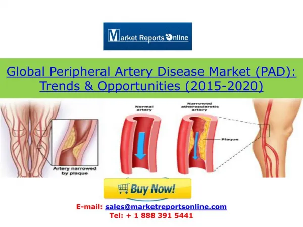
Emboli usually impact at branching points in arterial tree, particularly at the bifurcation of the aorta, the common femoral bifurcation & popliteal trifurcation. Sites of occlusion embloi to the lower limb: Femoral artery 45% Aorta & iliac artery 26% popliteal artery 15% tibial artery 1% WWW.SMSO.NET
Thrombosis: • Thrombosis usually occur on a pre-existing atherosclerotic lesion. • Occasionally thrombosis occur on relatively normal artery In patients with hypercoagulabale states ex: Pt with malignancy, polycythemia or pt taking high doses of oestrogen. Trauma • It is important to determine a history of arterial trauma, arterial catheterization, intra-arterial drug induced injection, limb fractures.
ClinicalFeatures • The6 P’s: ■ Pain. ■ Pallor. ■ Pulselessness. ■Perishing cold. ■ Paraesthesia. ■ Paralysis.
Clinical differentiation between thrombosis & embolism • Thrombosis: • No obvious cardiac source. • history of cluadication. • abnormal pulses in contralateral limb. • Angiogram: diffuse atherosclerotic • Well developed collateral Embolism: obvious cardiac source No hx of cluadication Normal pulses in contralateral limb Angiogram: minimal atherosclerotic Few collateral
TX:Immediately Anticoagulant with heparin to prevent propagation of thrombus & distal thrombosis & this achieved by giving a bolus of 10 000 units of heparin intravenously & an infusion of about 1000 units of heparin per hour
Example of acute arterial embolus “Saddle” Embolus of right iliac artery
Man Embolectomy :agement This operation usually performed under local anaesthesia. A groin incision is made & the common femoral artery is opened. often the clot is found in the artery a Fogarty balloon catheter is passed in turn into the proximal & distal arteries the balloon is inflated & the catheter withdrawn removing the clot. Fogarty balloon catheter
Management • Thrombolytic therapy: • Percutaneous intra-arterial thrombolytic therapy. Takes approximately 12-72 hours to dissolve the clot. • Agents used: streptokinase, urokinase & tissue plasminogen activator. • Mechanism: The convert plasminogen to plasmin which the active lytic agent.