Download
1 / 24
430 likes | 765 Vues
Arm and Elbow Anatomy Team 434. Color Index: I mportant Point s Helping notes Explanation. If you have any complaint or suggestion please don’t hesitate to contact us on: AnatomyTeam434@gmail.com. OBJECTIVES. Describe the attachments, actions and innervations of: Biceps brachii

E N D
Arm and Elbow Anatomy Team 434 Color Index: • Important Points • Helping notes • Explanation If you have any complaint or suggestion please don’t hesitate to contact us on: AnatomyTeam434@gmail.com
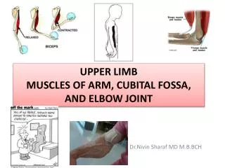
OBJECTIVES Describe the attachments, actions and innervations of: Biceps brachii Coracobrachialis Brachialis Triceps brachii Demonstrate the following features of the elbow joint: Articulating bones Capsule Lateral & medial collateral ligaments Synovial membrane Demonstrate the movements : flexion and extension of the elbow. List the main muscles producing the above movements. Define the boundaries of the cubital fossa and enumerate its contents
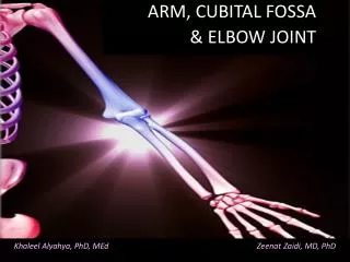
Introduction Shoulder Elbow The lateral and medial intermuscular septa divide the distal part of the arm into two compartments: -Anterior -Posterior Arm Humerus Elbow Anatomy This video explains the whole lecture!
Anterior Fascial Compartment Contents Muscles of the Anterior Compartment Coracobrachialis Muscles:Biceps brachii, Coracobrachialis &Brachialis. Blood Vessels: Brachial artery & Basilic vein. Nerves: Musculocutaneous, Median, Radial & Ulnar. Brachialis Bicepts trachi important notes: front muscles are 3 : biceps, coracobrachialis and brachialis that lies below them. posterior compartment contains triceps muscle.
Biceps Brachii Tendon inserted to radial tuberosity Biceps brachii Coracoid process Origin: Two heads: Long Head (lateral head) from supraglenoid tubercle of scapula(intracapsular) Short Head from the tip of coracoid process of scapula v The two heads join in the middle of the arm important notes: biceps muscle has two heads short and long (lateral) insertion: tendon to radial tuberosity into radius + to deep fascia of forearm. supination= lateral rotation of forearm. Insertion: in the posterior part of the radial tuberosity. into the deep fascia of the medial aspect of the forearm through bicipital aponeurosis. Nerve supply: Musculocutaneous Action: Strong supinator of the forearm, used in screwing. Powerful flexor of elbow Weak flexor of shoulder
Biceps Brachii Action • Strong supinator of the forearm, used in screwing. • Powerful flexor of elbow • Weak flexor of shoulder Biceps brachii Nerve supply Musculocutaneous Origin Insertion in the posterior part of the radial tuberosity. into the deep fascia of the medial aspect of the forearm through bicipital aponeurosis. Short Head Long Head from supraglenoid tubercle of scapula(intracapsular) from the tip of coracoid process of scapula The two heads join in the middle of the arm
Coracobrachialis Origin: Tip of the coracoid process of scapula (with short head of bicepes brachii ). Insertion: Middle of the medial side of the shaft of the humerus Nerve supply: Musculocutaneous Action: Flexor & a weak adductor of the arm
Brachialis Origin: Front of the lower half of humerus Insertion: Anterior surface of coronoid process of ulna Action: Strong flexor of the forearm Nerve supply: Musculocutaneous & Radial important notes: brachialis muscle has a double innervation form musculocutaneous and radial nerve. brachialis muscle continues to ulna.
Coracobrachialis & Brachialis Action Coracobrachialis Flexor & a week adductor of the arm Nerve supply Musculocutaneous Insertion Origin Middle of the medial side of the shaft of the humerus Tip of the coracoid process of scapula Action Strong flexor of the forearm Brachialis Nerve supply Musculocutaneous & Radial Insertion Origin Anterior surface of coronoid process of ulna Front of the lower half of humerus
Muscles of the Posterior Compartment Posterior Fascial Compartment Contents Triceps brachii Muscles: Triceps Vessels: Profunda brachii & Ulnar collateral arteries Nerves: Radial & Ulnar
Triceps Origin:Three heads: Long Head from infrglenoid tubercle of the scapula Lateral Head from the upper half of the posterior surface of the shaft of humerusabovethe spiral groove Medial Head from the lower half of the posterior surface of the shaft of humerus belowthe spiral groove Insertion: Common tendon inserted into the upper surface of the olecranon process of ulna Nerve supply: Radial nerve Action: Strong extensor of the elbow joint important notes: supraglenoid tubercle of scapula is intracapsular (inside capsule) long head’s origin is the infraglenoid tubercle. lateral and medial heads origins are on posterior surface of humerus. medial head is below radial groove in lower half . lateral head is above radial groove in upper half.
Triceps Action Strong extensor of the elbow joint Triceps Nerve supply Radial nerve Origin Insertion Common tendon inserted into the upper surface of the olecranon process of ulna Medial Head Lateral Head from the lower half of the posterior surface of the shaft of humerus belowthe spiral groove Long Head from the upper half of the posterior surface of the shaft of humerusabovethe spiral groove from infraglenoid tubercle of the scapula
Content of Cubital Fossa • Boundaries of Cubital Fossa Cubital Fossa • The Cubital fossa is a triangular depression on the anterior aspect of the elbow. • Cubital fossa has three boundries Boundaries of Cubital fossa Base: line through the two epicondyles of humerus Laterally : barachioadilis Medially: Pronator Teres important notes: brachial artery passes through cubital fossa ( that’s why we can measure blood pressure at cubital fossa) base is line between epicondyles. # Roof: Skin, superficial & deep fascia and bicipital aponeurosis # Floor: Brachialis medially and supinator laterally
Cubital Fossa Content of Cubital Fossa 1. Median nerve. 2. Brachial artery divides into radial & ulnar arteries. 3. Biceps brachii tendon. 4. Deep branch of radial nerve.
Elbow Joint • Elbow joint is sorted as Synovial Hinge joint . • The articular surfaces are covered by Hyaline cartilage. # Articulations: 1- Trochlea and capitulum of the humerus (Above) 2- Trochlea notch of ulna and the head of radius (Below) important notes: dislocation of elbow joint= posterior displacement of olecranon process from trochlea. dislocations occur mainly in children because their bones are less developed. avulsion=separation
Capsule important notes: behind medial epicondyle is ulnar nerve which is related directly to bone. the ulnar nerve is the largest unprotected nerve. anterior ligament limits extension.
Ligaments ( Lateral and Medial ) Features of the Lateral (Radial Collateral) Ligament: 1- Triangular in shape 2- Apex attached to lateral epicondyle of Humerus. 3- Base attached to the upper margin of annular ligament. important notes: annular ligament is around the head of radius and it connects radius with ulna. lateral ligament is triangular in shape. base is attached to annular ligament. medial ligament is very strong. ligament helps stability of joint. medial ligament is strong because of the anterior band.
Medial (Ulnar Collateral) Ligament composed of three bands : 1- Anterior strong cord-like band: Between medial epicondyle and the coronoid process of ulna. 2- Posterior weaker fan-like band: Between medial epicondyle and the olecranon process of ulna 3- Transverse band: Passes between the anterior and posterior bands.
Synovial Membrane Synovial Membrane does two functions : 1- lines the capsule. 2- covers the fatty acids pads in the floors of the coronoid,radial, and olecranon fossae. # Is continuous below with synovial membrane of the superior radio-ulnar joint. important notes: henge= movement around axis only. articular surfaces are covered by hyaline cartilages. capsule above humerus. synovial membrane of elbow continues down bursa=sac bursa= synovial membrane
Relations & Movement Relations: 1- Anterior: Brachialis, tendon of biceps, median nerve and brachialartery. 2- Posterior: Triceps muscle and Small bursa intervening. 3- Lateral: common extensor tendon and The supinator. 4- Medial: Ulnar nerve. The joint is supplied by branches from themedian, ulnar, musculocutaneous, and radial nerves. Movement:
Articulation & Applied Anatomy Carrying Angle # Carrying angle is the angle between the long axis of the extended forearm and the long axis of the arm. # it opens laterally. # it is 170 degrees in male and 167 degrees in females. # it disappears when the elbow joint is flexed. # it allows The forearms to clear the hips in swinging movements during walking and is important when carrying objects. important notes: the carrying angle opens laterally. male’s angle is larger than females’ angle disappears with flexion.
MCQ 7- Which of these joint is bounded three bones together:A- Elbow joint.B- Knee joint. C- Wrist joint.D- Shoulder joint. 8- The articular surface of elbow joint is covered with:A- Elastic cartilage.B- Hyaline cartilage.C- Fibrocartilage.D- Both B and C.9- The capsule attached to the margins of the of the coronoid process of the ulna from:A- Anteriorly above.B- Posteriorly above.C- Anteriorly below.D- Posteriorly below.10- Which band of the ulnar collateral ligaments is strong cord-like band:A- Anterior band.B- Posterior band.C- Transverse band.D- Annular ligament. 11- Which nerves is considered the largest unprotected nerve in the body:A- Median nerve.B- Radial nerve.C- Axillary nerve.D- Ulnar nerve. 8-B 9-C 10-A 11-D 1-Which of the following is presenting in the anterior fascial compartment:A- Musculocutaneous nerve.B- Ulnar nerve.C- Axillary nerve.D- Radial nerve.2- When the coracoid process has fractured, which muscle may affected:A- Pectoralis major.B- coracobrachialis.C- brachialis.D- None of the above.3- Which of these muscles related to anterior fascial compartment is supplied by radial nerve:A- Biceps brachii.B- coracobrachialis.C- Triceps D- Brachialis. 4- Muscle that Origin from the posterior surface of the humerus below the spiral groove:A- Medial head of triceps.B- Long head of triceps.C- Lateral head of triceps.D- B and C5- lateral boundary of the cubital fossa:A- Pronator teres.B- Two epicondyles of humerus.C- Brachioradialis.D- Brachialis. 6- Which of these content of cubital fossa of you cut it, you will lost abduction of thumb?A- Ulnar artery.B- Median nerve.C- Ulnar nerve.D- Radial artery. 4-A 5-C 6-B 7-A 1-A 2-B 3-D