Download
1 / 72
720 likes | 938 Vues
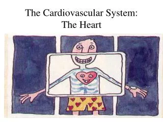
The Cardiovascular System: The Heart. Embryology of the Heart. Derived from mesoderm, begins development at 3 rd week of gestation Contraction of the heart begins by day 22 A pair of tubes develop (endothelial tubes), These fuse into the primitive heart atria and ventricles form

E N D
Embryology of the Heart • Derived from mesoderm, begins development at 3rd week of gestation • Contraction of the heart begins by day 22 • A pair of tubes develop (endothelial tubes), • These fuse into the primitive heart atria and ventricles form • These then assume a “U” shape then an “S” shape • Making “US”
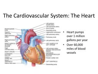
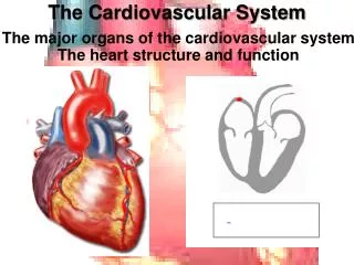
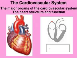
The Heart • Cardiovascular system • heart, arteries, veins and capillaries • 2 major divisions: • 1. Pulmonary circuit - right side of heart • carries deoxygenated blood to lungs • 2. Systemic circuit - left side of heart • supplies oxygenated blood to the body • Cardiology- study of the heart
Size, Shape and Position • Big a closed fist, males have bigger anatomical hearts • Located in the mediastinum, like a cone on its side between the lungs • Base - broad superior portion of heart • Apex - inferior end, tilts to the left, tapers to point
Pericardium • Pericardium- surrounds heart, keeps your heart in it’s place (like a father-in-law with the gun collection) • Allows heart to beat without friction, room to expand and resists excessive expansion • Pericarditis= inflammation of pericardium, can result in: • Cardiac Tamponade= fluid in the pericardial cavity compressing the heart, can stop the heart beat
Pericardium • Pericardium- Two parts: • 1.) Parietal Pericardium • Two layers: • Fibrous layer- superficial • protects, prevents overstretching, anchors heart • Serous layer- deep • 2.) Visceral Pericardium • Also called the epicardium
Heart Wall • 3 layers of the Heart wall • 1.) Epicardium- outside slippery layer • Also called the visceral pericardium (just to be confusing) • 2.) Myocardium- muscle of heart • 3.) Endocardium- inside the heart, covers chambers, heart valves • The heart can’t get oxygen from the inside. Why?
Heart Chambers • 4 chambers • Right and left atria (= entry halls) • 2 superior, posterior chambers • receive blood returning to heart • Right and left ventricles (= little bellies) • 2 inferior chambers • pump blood into arteries • Left ventricle is thicker, why?
Anterior Anatomy – Atrioventricular sulcus - separates atria, ventriclesAnterior and posterior interventicular sulci - grooves separate ventricles Atrioventricular sulcus
Posterior Heart AnatomyPosterior Interventricular Sulcus- groove separating ventricles
Heart Chambers - Internal • Interatrial septum • wall that separates atria • Pectinate muscles • internal ridges of myocardium in right atrium and both auricles • Interventricular septum • wall that separates ventricles • Trabeculae carneae • internal ridges in both ventricles walls • Chordae tendineae- cords connecting to the tricuspid and mitral valves
Heart Valves • Ensure one-way blood flow • Semilunar valves - control flow into great arteries • pulmonary: from right ventricle into pulmonary trunk • aortic: from left ventricle into aorta • Atrioventricular (AV) valves • right AV valve has 3 cusps (tricuspid valve) • left AV valve has 2 cusps (mitral,bicuspid valve) • rat lamb • chordae tendineae - cords connect AV valves to papillary muscles (on floor of ventricles)
Valve Anatomy • The AV valves, the tricuspid and bicuspid (mitral) valves
AV Valve Mechanics • Ventricles relax, pressure drops, semilunar valves close, AV valves open, blood flows from atria to ventricles • Ventricles contract, AV valves close(papillary m. contract and pull on chordae tendineae to prevent prolapse),pressure rises, semilunar valves open, blood flows into great vessels
Be the Blood - Discretion I • You (blood) flow into heart in the Superior (S) vena cava and inferior (I) vena cava and then into the right atrium (1) • You pass through the tricuspid (= three points) valves (2) into the right ventricle (3) • You go through the pulmonary semilunar (4) valves into the pulmonary trunk (5) • The pulmonary trunk divides into right and left pulmonary arteries (6) which go to the lungs so that you can be oxygenated
Be the Blood II • You (be the blood!) then flow from the lungs through the pulmonary veins (7) into the left atrium (8) • Through the bicuspid valve (9) to the left ventricle (10) • The left ventricle pumps blood (you) through the aortic semilunar valve (11) into the ascending aorta (12) then to the aortic arch and on to the descending aorta • http://www.youtube.com/watch?v=upctPUa6RhA
Circulation to the Heart • The heart can not get nutrients from inside • Blood vessels must supply the heart muscle this is called the coronary circulation • The arteries of the heart encircle it like a crown • While contacted no blood flows to the heart • When the heart relaxes the blood flows to it • FLOW- Aorta to coronary arteries to coronary veins
Heart Valves 2 ? 1?
Coronary Circulation • Left coronary artery • 1. anterior interventricular artery • supplies interventricular septum + anterior walls of ventricles • 2. circumflex artery • passes around left side of heart in coronary sulcus, supplies left atrium and posterior wall of left ventricle • Right coronary artery • 1. posterior interventricular artery • supplies posterior walls of ventricles • 2. marginal artery • supplies lateral R atrium + ventricle
Coronary Circulation memory aid • Remember: a heart that doesn’t work well will often- LAC RPM • Right Coronary artery gives off the- • Right Posterior Interventricular artery and Marginal artery (RPM) • Left Coronary artery gives off the- • Left Anterior Interventricular artery and the Circumflex artery (LAC)
Structure of Cardiac Muscle • Compared to skeletal muscle cardiac muscle is: • Short in length • Branched • One to two nuclei • Has intercalated discs, these contain gap junctions that assist in electrical conduction
Metabolism of Cardiac Muscle • Aerobic respiration • Rich in myoglobin and glycogen • Large mitochondria • Organic fuels: fatty acids, glucose, ketones • What should our diets be? • Fatigue resistant
Heart Electrical Conduction • Myogenic - heartbeat originates within the heart • The fundamental rhythm of the heart is automatic and not dependent on the nervous system or endocrine system • This rhythm is due to autorhythmic cells, specialized cardiac muscle fibers that are self-exciting • These cells act as: a Pacemaker… • Setting the rhythm of the entire heart • …and Conduction system • A route for conducting electrical energy throughout the heart muscle
It’s All In The Timing • The chambers of the heart have to be coordinated or the heart will not work • The SA node (sinoatrial node) stimulated first, as the name implies this node causes atria contraction • Next the AV node (atrioventricular node) is stimulated • The impulse then goes to the Bundle of His (Atrioventricular bundle) • The right and left bundle branches goes to the apex of the heart • Finally, conduction myofibers termination is called Purkinje fibers that conduct impulses to the rest of the heart
Cardiac Conduction System Bundle of His
Heart Conduction: Memory Aid • Remember: SAVe HIS Right and Left BUN from PURKing
Cardiac Rhythm • Systole = contraction; diastole = relaxation • Sinus rhythm • set by SA node, adult at rest is 70 to 80 bpm • Ectopic foci - region of spontaneous firing (not SA) • nodal rhythm - set by AV node, 40 to 50 bpm • intrinsic ventricular rhythm - 20 to 40 bpm • Arrhythmia - abnormal cardiac rhythm • heart block: failure of conduction system • bundle branch block • total heart block (damage to AV node)
Electrocardiogram (ECG) • Composite of all action potentials of nodal and myocardial cells detected, amplified and recorded by electrodes on arms, legs and chest
ECG Alphabet • The ECG alphabet goes PQRST • P wave- the first wave shown • QRS complex- the middle quibble • T wave- the last wave
Pacemakers • The SA node is the pacemaker of the heart • If the SA node becomes diseased, and heart rate gets too low (<40 BPM) an Artificial Pacemaker can be surgically implanted • Other sites besides the SA node can stimulate the heart, this is called: an Ectopic pacemaker • Triggers of this activity are caffeine, nicotine, that special someone
EKG/ ECG • Cardiac muscle contraction is electrical, the physiology is similar to skeletal muscle (see A&PI) these electrical impulses can be detected on the skin of the chest • Our friend the Aardvark! • This recording is called is called an Electrocardiogram and the machine is called an Electrocardiograph • The ECG tells three things: E- electrical conduction problems in the heart, C- cardiac enlargement, G- Cardiac Damage
Waves of Meaning I • First wave= P wave- atrial Depolarization • Second wave= QRS- ventricular depolarization as the electrical excitement spreads through the ventricles • Repolarization of the atria occurs here but is not seen due to the QRS length • Third wave= T wave- ventricular REploarization
ECG/EKG • Abnormal Waves • Larger P= enlarged atrium • Enlarged R= enlarged ventricles
EKG (ECG) • Time span between waves is called intervals or segments • P-Q interval lengthened- coronary artery disease, rheumatic fever • Q-T interval lengthened- myocardial damage, HEART ATTACK, coronary ischemia or conduction anomalies
ECGs, Normal & Abnormal No P waves
ECGs, Abnormal Extra systole: note the inverted QRS complex, misshapen QRS and T and absence of a P wave preceding this contraction.
ECGs, Abnormal Arrhythmia: conduction failure at AV node No pumping action occurs
Cardiac Cycle • Events in one heartbeat • Systole is contraction, Diastole is relaxation (“Relax? Relax? When I die, then I’ll relax.”) • The cycle consists of systole and diastole of both atria plus systole and diastole of both ventricles
Heart Sounds • Listening to the sounds of the body is called auscultation (= listening), you can hear bowel sounds, breathing, and heart beats with your ear to the patient (old method) or with a stethoscope (modern method) • 4 heart sounds, 2 clinically important (because a stethoscope can hear them): • The first sound is lubb, • First heart sound (S1), louder and longer “lubb”, occurs with closure of AV valves • The second sound is dub • Second heart sound (S2), softer and sharper “dub” occurs with closure of semilunar valves
Lubb- Dub • Lubb- closure of AV valves: • Tricuspid valve • Mitral valve • Dub- closure of the semilunar valves: • Aortic • Pulmonary Dub Dub Lubb Lubb