Download
1 / 40
400 likes | 413 Vues
This article provides an overview of the different structures and functions of the eye, including the orbit, extrinsic eye muscles, optic nerves, visual accessory organs, outer tunic, middle tunic, vascular tunic, inner tunic, retina, photoreceptors, light refraction, and common vision problems. It also includes a video of a real LASIK surgery.

E N D
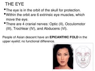
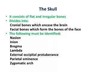
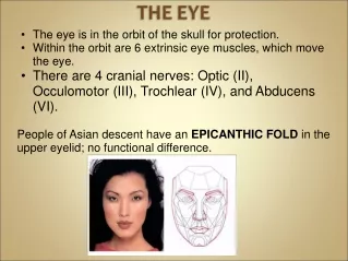
The eye is in the orbit of the skull for protection. • Within the orbit are 6 extrinsic eye muscles, which move the eye. • There are 4 cranial nerves: Optic (II), Occulomotor (III), Trochlear (IV), and Abducens (VI). • People of Asian descent have an EPICANTHIC FOLD in the upper eyelid; no functional difference.
Visual Accessory Organs • Eyelid • Conjuctiva • Lacrimal Gland • Extrinsic Muscles
Eyelid • Covers and protects the eye, thin skin • Skin will not protect you from intense radiation, that’s why we use special goggles in a tanning bed
CONJUNCTIVA is like a covering around the eye and under the eyelids. • PINK EYE (layman’s term), known as CONJUNCTIVITIS (from bacteria, very contagious).
Extrinsic Eye Muscles Moves the eyeball
LACRIMAL GLANDS are the largest set. They are on the superior lateral eyelid and they produce tears, which drain into the nasal cavity via the LACRIMAL DUCT. • The function is to moisten and lubricate the eye surface, and it has enzymes to kill bacteria (which thrive in warm, moist conditions).
Outer Tunic • Cornea - transparent, focuses light rays • Sclera – continuation of cornea, going toward the back of the eye (white of the eye) • Optic Nerve – exits at the optic disk and transmits visual information from the eye to the brain.
Middle Tunic Choroid Coat – contains blood vessels Ciliary Body – holds the lens in place Lens – focusing Iris – colored portion of the eye Aqueous humor – liquid surrounding the lens Pupil – opening for light to enter
The Vascular Tunic PLAY Vascular Tunic (Uvea) Figure 16.8
Retina - visual receptor cells Fovea Centralis - region of the sharpest vision (aka, macula) Optic Disc – where nerve fibers leave the eye, creating the blind spot Vitreous Humor – supports internal parts, fluid Inner Tunic
Retina The retina is made up of PHOTORECEPTORS, which are sensors for light.
Rods = monochromatic (b&w) Cones = color vision
PhotoreceptorsRods & Cones Figure 16.11
Light Refraction Light bends around objects Images viewed by the eye are upside down
R.O.Y.G.B.I.V Rainbows are seen after rain because light is passing through water droplets. This separates the white light into the individual colors of the spectrum
The Eye as an Optical Device Figure 16.14a–c
We have difficult interpreting images that are upside down Which one is the real mona lisa?
Fun Fact: -When you are looking at someone you love, your pupils dilate, and they do the same when you are looking at someone you hate.
Problems with the Eyes Clouding of the lens leads to a clinical condition known asCATARACTS. Treatment is to remove the lens and replace it with a plastic one (which is not flexible either).
Problems with the IRIS and PUPIL The function is to constrict or dilate the pupil (opening) to allow light in. Therefore, it regulates the amount of light passing to the visual receptors of the eye. ANIRIDIA = a condition where a person is born without an iris
Why are all babies born with blue eyes? • Melanin is a brownish pigment that adds color to your hair, eyes, and skin. At the time babies are born, melanin hasn't yet been "deposited" in the eyes' iris. Hence, they appear blue. • After about six months, eyes change color depending on the amount of melanin. If you have a lot of it, your eyes will turn dark brown. If you have little, they'll stay blue. And if you have no melanin, your eyes may appear pink (albino). .
A genetic trait that affects boys more than girls. The location of the gene is on the X chromosome Colorblindness
The region where the optic nerve and blood vessels goes in and out of the eye has no photoreceptors = BLIND SPOT. • Hold your hands out at 45° and that’s the location of the blind spot. • You can still see your hands because the other eye sees it. Close your right eye and look for your right hand and you’ll find the blind spot.
FLOATERS are when a capillary breaks and cells break off. Floaters don’t actually move, the eye just tries to track them.
HYPEROPIA(far-sighted)eyes are too short MYOPIA(nearsighted)eyes are too long
ASTIGMATISM ASTIGMATISM is when the cornea has an irregular shape. Part of the field of view is out of focus. They eyeball changes shape until age 24.